Chapter 3. Biopsychology

3.2 Cells of the Nervous System
Biological psychologists striving to understand the mind and behavior, study the nervous system in various ways. The nervous system is an electro-chemical communication system, which receives and processes information from the outside world, and allows us to react to it. The nervous system is composed of two basic cell types: neurons and glial cells. The neurons (nerve cells) are the central building blocks of the nervous system, whereas various types of glial cells provide support services.
Neuron Structure
Like all cells, neurons consist of several different parts, each serving a specialized function ( Figure 3.4 ). The cell body or soma contains the nucleus (which contains the genetic material of the cell), and other structures that are important for cell maintenance. The soma is surrounded by a semipermeable membrane, which regulates what can pass in and out of the neuron. Information enters the neuron through the dendrites, which are branching tree-like projections on the soma. Information is then transmitted to the other end of the neuron down a tail-like process called the axon. Information travels through the neuron in the form of electrical energy. At the end of the axon, there are small structures called terminal buttons which contain chemical messengers called neurotransmitters; these allow the neuron to communicate with other cells.

The length of the axon determines how far one neuron carries information through our bodies, some axons are microscopic while others, like the ones running from our toes to the base of our spine, are several feet long. Many axons are coated with a fatty substance known as the myelin sheath , which acts as an insulator and speeds up the transmission of the electrical signals down the axon. The myelin has small gaps along the axon called the nodes of Ranvier. In myelinated axons, the electrical signals are only generated at the nodes, like a train making express stops. In unmyelinated neurons, information travels more slowly because electrical signals need to be generated along the entire length of the axon—acting more like a local train. Multiple sclerosis (MS) is an autoimmune disorder that causes degeneration of the myelin sheath on axons throughout the nervous system. This can result in inefficient transmission, and sometimes total loss, of neural information. Some of the first symptoms of MS include blurry vision, sensations of numbness (like pins and needles) and difficulty walking.
Link to Learning
Watch this video about neuronal communication to learn more.
Neuronal Communication
Now that we have learned about the basic structures of the neuron, let’s take a closer look at the signals that the neurons transmit. We will look first at how the electrical signals are generated and move through the neuron. Then we will look at how the chemical messengers are released and communicate with other cells, including other neurons.
The neuron exists in a fluid environment—it is surrounded by extracellular fluid and contains intracellular fluid. The neuronal membrane keeps these two salty fluids separate. Both fluids contain sodium (Na+) and potassium (K+) ions, and other charged molecules. Differences in the overall electrical charge of molecules in the two fluids contribute to a difference in voltage across the membrane, called the membrane potential. When the neuron is resting, the membrane resting potential is about 70 mV and the inside of the neuron is more negatively charged than the outside. In general, ions move quickly across concentration gradients from areas of high concentration to areas of low concentration through the process of osmosis. Na + is at higher concentrations outside the neuron, so it strongly wants to move into the neuron. K + on the other hand, is more concentrated inside the neuron, and so wants to move out of the neuron (Figure 3.5). Because the inside of the neuron is slightly negatively charged compared to the outside, this provides an additional drive for Na+ to be drawn into the neuron (opposite electrical charges attract). We call this an electrical gradient. However, there is relatively little ionic movement across the membrane when the neuron is resting. This is because most of the Na+ and K+ gates in the semi-permeable membrane, which regulate the flow of ions, are closed when the neuron is at rest. This results in a high state of tension across the membrane; the Na+ and K+ ions are waiting for the gates to open so they can move through them, and so the neuron is primed and ready for action. For the most part, the Na+ and K+ gates do a good job of staying closed, but a few open and there is a little bit of leakage of Na+ and K+ in the direction described above. To keep the resting potential stable, there is an active sodium-potassium pump, which pumps three sodium ions out of the neuron for every two potassium ions in.

From this resting potential state, if the neuron receives a signal at the dendrites (Figure 3.5), the Na+ gates open on the neuronal membrane, and Na+ ions rush into the cell. These ions make the inside of the cell become more positively charged. If that charge reaches a certain level, called the threshold of excitation, the neuron becomes active and the action potential begins. The action potential, sometimes called a nerve impulse, is a large, rapid change in voltage across the membrane. (Figure 3.6).

Whether or not the threshold of excitation is reached depends on the strength of the signal being received. When the signal is strong, more Na+ gates open and more Na+ ions enter and the inside of the cell becomes more positive. When the threshold of excitation is reached, all the Na+ channels open rapidly, causing a massive influx of Na + ions and a huge positive spike in the membrane potential. At the peak of the spike, the Na+ gates close and the K+ gates open. As positively charged potassium ions rapidly leave the neuron, the inside of the cell quickly starts to become less positive (or more negative). This process is called repolarization. After an action potential is generated there is a resting period, called the refractory period, where the inside of the cell is in a state of hyperpolarization. This means that it is slightly more negative than the resting potential for a few milliseconds; it is not possible to generate another action potential during the refractory period. As the K+ gates close, the neuron returns to the resting potential (Figure 3.6).
The action potentials are generated in the cell body first and then are produced along the axon until they reach its end. At each point in the journey, some of the sodium ions that enter the neuron diffuse to the next section of the axon, raising the charge past the threshold of excitation and triggering a new influx of sodium ions. So, multiple action potentials are generated sequentially all the way down the axon until they reach the terminal buttons.
The action potential is an all-or-none phenomenon. In simple terms, this means that an incoming signal from another neuron must be strong enough to reach the threshold of excitation, otherwise, there is no action potential (none). The action potential is always generated at its full strength at every point along the axon. It does not fade away as it travels down the axon. This ensures that there is no loss of information along the way. The size of the action potential does not depend on the strength of the stimulus, it is always the same. Strong stimuli simply produce more action potentials.
In many ways, the way that a neuron produces action potentials is similar to the way that an old fashioned toilet produces a flush. Both work on the all or none principle. A toilet also has a threshold of excitation – if you do not push the handle hard enough, it will not flush. However, as long as you press the handle hard enough, the toilet always produces a flush of the same size. So, you have all or none. If you push very hard, you don’t get any more water than if you push more gently, provided you meet the threshold of excitation. Like a neuron, a toilet also has a refractory period (when the tank is refilling); the toilet cannot flush during this time.
Communication across the synapse
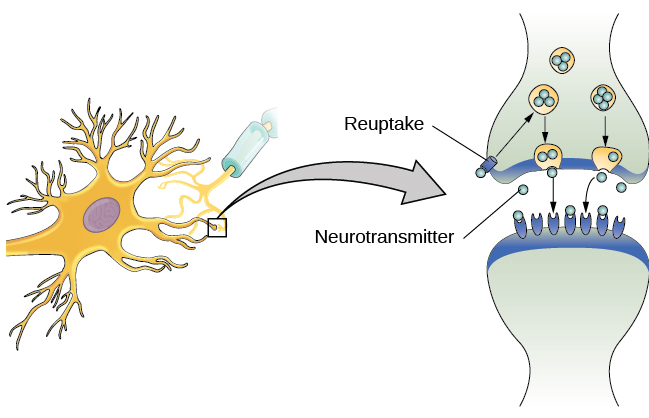
At the end of every axon, there is a tiny gap called a synapse between the terminal buttons and the next cell, which is often another neuron. This gap is filled with extracellular fluid and is too big for an action potential to jump across. Instead, when the action potential arrives at the terminal button, small vesicles release their neurotransmitters into the synapse. The neurotransmitters travel across the synapse and bind to receptors on the dendrites of the adjacent neuron. This action then affects the ionic gates in that neuron. Some neurotransmitters are excitatory (they open Na+) gates, whereas others are inhibitory (they open K+) gates. So, neurotransmitters give more flexibility in terms of how neurons can respond to a stimulus.
Neurotransmitters and Drugs
There are several different types of neurotransmitters in our nervous system, each one acts on specific types of receptors and has a particular action (see Table 3.1 ).
Table 3.1 Major Neurotransmitters and How They Affect Behavior
| Acetylcholine | Muscle action, memory | Increased arousal, enhanced cognition |
| Beta-endorphin | Pain, pleasure | Decreased anxiety, decreased tension |
| Dopamine | Mood, sleep, learning | Increased pleasure, suppressed appetite |
| Gamma-aminobutyric acid (GABA) | Brain function, sleep | Decreased anxiety, decreased tension |
| Glutamate | Memory, learning | Increased learning, enhanced memory |
| Norepinephrine | Heart, intestines, alertness | Increased arousal, suppressed appetite |
| Serotonin | Mood, sleep | Modulated mood, suppressed appetite |
Much of what psychologists know about the functions of neurotransmitters comes from research on the effects of drugs in psychological disorders. There is some biological evidence that psychological disorders like depression and schizophrenia are associated with imbalances in one or more neurotransmitter systems. Psychoactive medications affect neurotransmitter balance and can help improve the symptoms associated with these disorders.
Psychoactive drugs generally act as agonists or antagonists for a given neurotransmitter system, which is why they can affect our behaviors, thoughts, and feelings. Agonists are chemicals that mimic the action of a neurotransmitter at the receptor site. An antagonist, on the other hand, blocks or impedes the normal activity of a neurotransmitter at the receptor. GABA-agonists such as benzodiazepines mimic the effects of GABA and so produce calming effects for people who have anxiety. Conversely, psychotic symptoms in schizophrenia are associated with high levels of dopamine and so are treated with dopamine antagonists. These drugs stop dopamine’s effects by blocking its receptors. Thus, they can prevent one dopamine-producing neuron from sending information to adjacent neurons.
In contrast to agonists and antagonists, which both operate by binding to receptor sites, some psychoactive drugs affect the deactivation of the neurotransmitter. Once the neurotransmitter has done its job it is typically removed in some way so that it does not continue to act on the post-synaptic neuron. In some cases, the neurotransmitters in the synapse diffuse away, or they are broken down by an enzyme or pumped back into the pre-synaptic neuron in a process known as reuptake ( Figure 3. 7 ).

Some psychoactive drugs can prevent the removal of the neurotransmitters. This allows neurotransmitters to remain active in the synapse for longer durations, increasing their effectiveness. Depression, which has been linked with reduced serotonin levels, is frequently treated with selective serotonin reuptake inhibitors (SSRIs). By preventing reuptake, SSRIs prolong the action of serotonin, giving it more time to interact with serotonin receptors on dendrites. So, SSRIs act as serotonin agonists, even though they do not work directly on the receptors. Common SSRIs include Prozac, Paxil, and Zoloft. Psychotropic drugs are not instant solutions for people suffering from psychological disorders. Often, an individual must take a drug for several weeks before seeing improvement, and many psychoactive drugs have significant negative side effects. Furthermore, individuals vary dramatically in how they respond to the drugs. To improve chances for success, it is common for people receiving medications to undergo psychological and/or behavioral therapies as well. Research suggests that combining drug therapy with other forms of therapy tends to be more effective than any one treatment alone for the treatment of depression (Cuijpers, Noma, et al., 2020) and some anxiety disorders like panic disorder and obsessive-compulsive disorder (Cuijpers, Sijbrandij, et al., 2020).
Introduction to Psychology (A critical approach) Copyright © 2023 by Jill Grose-Fifer; Rose M. Spielman; Kathryn Dumper; William Jenkins; Arlene Lacombe; Marilyn Lovett; and Marion Perlmutter is licensed under a Creative Commons Attribution-NonCommercial 4.0 International License , except where otherwise noted.

Share This Book
Review Questions
Which of the following cavities contains a component of the central nervous system?
Which structure predominates in the white matter of the brain?
- myelinated axons
- neuronal cell bodies
- ganglia of the parasympathetic nerves
- bundles of dendrites from the enteric nervous system
Which part of a neuron transmits an electrical signal to a target cell?
Which term describes a bundle of axons in the peripheral nervous system?
Which functional division of the nervous system would be responsible for the physiological changes seen during exercise (e.g., increased heart rate and sweating)?
What type of glial cell provides myelin for the axons in a tract?
- oligodendrocyte
- Schwann cell
- satellite cell
Which part of a neuron contains the nucleus?
- synaptic end bulb
Which of the following substances is least able to cross the blood-brain barrier?
- sodium ions
- white blood cells
What type of glial cell is the resident macrophage behind the blood-brain barrier?
What two types of macromolecules are the main components of myelin?
- carbohydrates and lipids
- proteins and nucleic acids
- lipids and proteins
- carbohydrates and nucleic acids
If a thermoreceptor is sensitive to temperature sensations, what would a chemoreceptor be sensitive to?
Which of these locations is where the greatest level of integration is taking place in the example of testing the temperature of the shower?
- skeletal muscle
- spinal cord
- cerebral cortex
How long does all the signaling through the sensory pathway, within the central nervous system, and through the motor command pathway take?
- 1 to 2 minutes
- 1 to 2 seconds
- fraction of a second
- varies with graded potential
What is the target of an upper motor neuron?
- lower motor neuron
What ion enters a neuron causing depolarization of the cell membrane?
Voltage-gated Na + channels open upon reaching what state?
- resting potential
- repolarization
What does a ligand-gated channel require in order to open?
- increase in concentration of Na + ions
- binding of a neurotransmitter
- increase in concentration of K + ions
- depolarization of the membrane
What does a mechanically gated channel respond to?
- physical stimulus
- chemical stimulus
- increase in resistance
- decrease in resistance
Which of the following voltages would most likely be measured during the relative refractory period?
Which of the following is probably going to propagate an action potential fastest?
- a thin, unmyelinated axon
- a thin, myelinated axon
- a thick, unmyelinated axon
- a thick, myelinated axon
How much of a change in the membrane potential is necessary for the summation of postsynaptic potentials to result in an action potential being generated?
A channel opens on a postsynaptic membrane that causes a negative ion to enter the cell. What type of graded potential is this?
- depolarizing
- repolarizing
- hyperpolarizing
- non-polarizing
What neurotransmitter is released at the neuromuscular junction?
- norepinephrine
- acetylcholine
What type of receptor requires an effector protein to initiate a signal?
- biogenic amine
- ionotropic receptor
- cholinergic system
- metabotropic receptor
Which of the following neurotransmitters is associated with inhibition exclusively?
This book may not be used in the training of large language models or otherwise be ingested into large language models or generative AI offerings without OpenStax's permission.
Want to cite, share, or modify this book? This book uses the Creative Commons Attribution License and you must attribute OpenStax.
Access for free at https://openstax.org/books/anatomy-and-physiology/pages/1-introduction
- Authors: J. Gordon Betts, Kelly A. Young, James A. Wise, Eddie Johnson, Brandon Poe, Dean H. Kruse, Oksana Korol, Jody E. Johnson, Mark Womble, Peter DeSaix
- Publisher/website: OpenStax
- Book title: Anatomy and Physiology
- Publication date: Apr 25, 2013
- Location: Houston, Texas
- Book URL: https://openstax.org/books/anatomy-and-physiology/pages/1-introduction
- Section URL: https://openstax.org/books/anatomy-and-physiology/pages/12-review-questions
© Jan 27, 2022 OpenStax. Textbook content produced by OpenStax is licensed under a Creative Commons Attribution License . The OpenStax name, OpenStax logo, OpenStax book covers, OpenStax CNX name, and OpenStax CNX logo are not subject to the Creative Commons license and may not be reproduced without the prior and express written consent of Rice University.

12.3 The Function of Nervous Tissue
Learning objectives.
By the end of this section, you will be able to:
- Describe the pathway involved with neural sensation, integration and motor response.
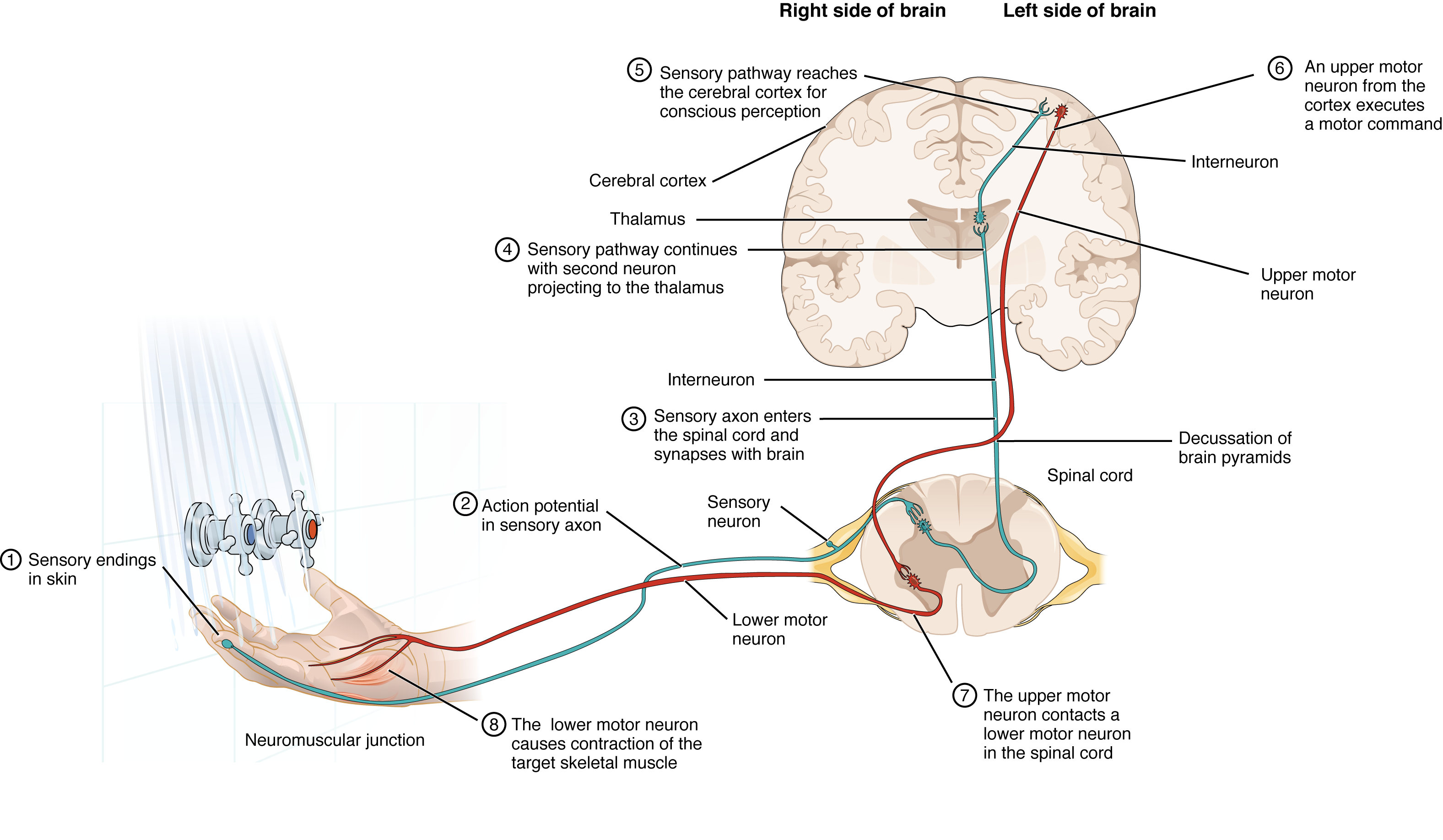
Having looked at the components of nervous tissue, and the basic anatomy of the nervous system, next comes an understanding of how nervous tissue is capable of communicating within the nervous system. Before getting to the nuts and bolts of how this works, an illustration of how the components come together will be helpful. An example is summarized in Figure 12.3.1 .

Imagine you are about to take a shower in the morning before going to school. You have turned on the faucet to start the water as you prepare to get in the shower. You put your hand out into the spray of water to test the temperature. What happens next depends on how your nervous system interacts with the stimulus of the water temperature and what you do in response to that stimulus.
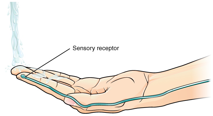
Found in the skin is a type of sensory receptor that is sensitive to temperature, called a thermoreceptor . When you place your hand under the shower (1 in Figure 12.3.1 , close up in Figure 12.3.2 ), the cell membrane of the thermoreceptors changes its electrical state (voltage). The amount of change is dependent on the strength of the stimulus (in this example, how hot the water is). This is called a graded potential . If the stimulus is strong, the voltage of the cell membrane will change enough to generate an electrical signal that will travel down the axon. The voltage at which such a signal is generated is called the threshold , and the resulting electrical signal is called an action potential . In this example, the action potential travels—a process known as propagation—along the axon from the initial segment found near the receptor to the axon terminals and into the synaptic end bulbs in the central nervous system (2 in Figure 12.3.1 ). When this signal reaches the end bulbs, it causes the release of a signaling molecule called a neurotransmitter .

In the central nervous system (in this case, the spinal cord), the neurotransmitter diffuses across the short distance of the synapse and binds to a receptor protein of the target neuron. When the neurotransmitter binds to the receptor, the cell membrane of the target neuron changes its electrical state and a new graded potential begins. If that graded potential is strong enough to reach threshold, the second neuron generates an action potential at its initial segment f that graded potential is strong enough to reach threshold, the second neuron generates an action potential at its initial segment (3 in Figure 12.3.1 ). The target of this neuron is another neuron in the thalamus of the brain, the part of the CNS that acts as a relay for sensory information. At this synapse, neurotransmitter is released and binds to its receptor. The thalamus then sends the sensory information to the cerebral cortex , the outermost layer of gray matter in the brain, where conscious perception of that water temperature begins.
Within the cerebral cortex, information is processed among many neurons, integrating the stimulus of the water temperature with other sensory stimuli, as well as with your emotional state and memories. Finally, a plan is developed about what to do, whether that is to turn the temperature up, turn the whole shower off and go back to bed, or step into the shower. To do any of these things, the cerebral cortex has to send a command out to your body to move muscles.
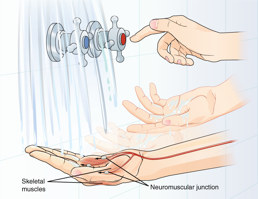
A region of the cortex is specialized for sending signals down to the spinal cord for movement. The upper motor neuron starts in this region, called the precentral gyrus of the frontal cortex , and has an axon that extends all the way down the spinal cord. The upper motor neuron synapses in the spinal cord with a lower motor neuron, which directly stimulates muscle fibers to contract. In the manner described in the chapter on muscle tissue, an action potential travels along the motor neuron axon into the periphery. The lower motor neuron axon terminates on muscle fibers at the neuromuscular junction. Acetylcholine is the neurotransmitter released at this specialized synapse, and binding to receptors on the muscle cell membrane causes the muscle action potential to begin. When the lower motor neuron excites the muscle fiber, the muscle contracts ( Figure 12.3.3 ). All of this occurs in a fraction of a second, but this story is the basis of how the nervous system functions.

Career Connections – Neurophysiologist
There are many pathways to becoming a neurophysiologist. One path is to become a research scientist at an academic institution. A Bachelor’s degree will get you started, and for neurophysiology that might be in biology, psychology, computer science, engineering, or neuroscience. But the real specialization comes in graduate school. There are many different programs out there to study the nervous system, not just neuroscience itself. Most graduate programs are doctoral, and are usually considered five-year programs, with the first two years dedicated to course work and finding a research mentor, and the last three years dedicated to finding a research topic and pursuing that with a near single-mindedness. The research will usually result in a few publications in scientific journals, which will make up the bulk of a doctoral dissertation. After graduating with a Ph.D., researchers will go on to find specialized work called a postdoctoral fellowship within established labs. In this position, a researcher starts to establish their own research career with the hopes of finding an academic position at a research university.
Other options are available if you are interested in how the nervous system works. Especially for neurophysiology, a medical degree might be more suitable so you can learn about the clinical applications of neurophysiology. Biotechnology firms are eager to find motivated scientists ready to tackle the tough questions about how the nervous system works so that therapeutic chemicals can be tested on some of the most challenging disorders such as Alzheimer’s disease or Parkinson’s disease, or spinal cord injury.
Others with a medical degree and a specialization in neuroscience go on to work directly with patients, diagnosing and treating mental disorders. You can do this as a psychiatrist, a neuropsychologist, a neuroscience nurse, or a neurodiagnostic technician, among other possible career paths.
Chapter Review
Sensation starts with the activation of a sensory receptor, such as the thermoreceptor in the skin sensing the temperature of the water. The sensory receptor in the skin initiates an electrical signal that travels along a sensory axon within a nerve into the spinal cord, where it synapses with a neuron in the gray matter of the spinal cord. At the synapse the temperature information represented in that electrical signal is passed to the next neuron by a chemical signal (the neurotransmitter) that diffuses across the small gap of the synapse and initiates a new electrical signal. That signal travels through the sensory pathway to the brain, synapsing in the thalamus, and finally the cerebral cortex where conscious perception of the water temperature occurs. Following integration of that information with other cognitive processes and sensory information, the brain sends a command back down to the spinal cord to initiate a motor response by controlling a skeletal muscle. The motor pathway is composed of two cells, the upper motor neuron and the lower motor neuron. The upper motor neuron has its cell body in the cerebral cortex and synapses with the lower motor neuron in the gray matter of the spinal cord. The axon of the lower motor neuron extends into the periphery where it synapses with a skeletal muscle fiber at a neuromuscular junction.
Review Questions
Critical thinking questions.
1. Suppose the thalamus were damaged at the area where the second sensory neuron synapsed with the third sensory neuron. Would you be able to consciously feel the water temperature? Why or why not?
2. Suppose the upper motor neuron were damaged. What symptoms would you expect?
Answers for Critical Thinking Questions
- If the thalamus were damaged at the site of synapsing between the second sensory neuron and the third sensory neuron, signals would not reach the cerebral cortex. This would result in a person not being able to detect the temperature information consciously.
- If the upper motor neuron were damaged, you would expect that someone would not be able to activate the lower motor neuron and then the muscle innervated by that lower motor neuron would not be able to move when brain signals asked for it to move. However, the lower motor neuron would be able to participate in.
This work, Anatomy & Physiology, is adapted from Anatomy & Physiology by OpenStax , licensed under CC BY . This edition, with revised content and artwork, is licensed under CC BY-SA except where otherwise noted.
Images, from Anatomy & Physiology by OpenStax , are licensed under CC BY except where otherwise noted.
Access the original for free at https://openstax.org/books/anatomy-and-physiology/pages/1-introduction .
Anatomy & Physiology Copyright © 2019 by Lindsay M. Biga, Staci Bronson, Sierra Dawson, Amy Harwell, Robin Hopkins, Joel Kaufmann, Mike LeMaster, Philip Matern, Katie Morrison-Graham, Kristen Oja, Devon Quick, Jon Runyeon, OSU OERU, and OpenStax is licensed under a Creative Commons Attribution-ShareAlike 4.0 International License , except where otherwise noted.

IMAGES
VIDEO
COMMENTS
Neuron - Critical Thinking Questions Flashcards | Quizlet. How does botulism toxin produce difficulty swallowing & breathing? Click the card to flip 👆. it binds to presynaptic vesicles and prevents the release of acetylcholine. Thus action potentials in nerve cells cannot produce them in skeletal muscles and they become paralyzed.
Critical Thinking Questions; Regulation, Integration, and Control. 12 The Nervous System and Nervous Tissue. ... Which type of neuron, based on its shape, is best ...
Study with Quizlet and memorize flashcards containing terms like 1) Draw and label the three parts of the neuron and explain the function of the dendrite and axon, 2) Name the three types of neurons classified according to the directions in which the impulse is transmitted.
Ch. 26 Critical Thinking Questions - Biology for AP® Courses | OpenStax. Highlights. 9. When you stick your hand in a bucket of ice, it grows numb after a while. Based on what you know regarding neuronal signaling, explain how the sensation of touch is blocked from signaling to the brain. 10.
The nervous system is composed of two basic cell types: neurons and glial cells. The neurons (nerve cells) are the central building blocks of the nervous system, whereas various types of glial cells provide support services.
Critical Thinking Questions 1. If a postsynaptic cell has synapses from five different cells, and three cause EPSPs and two of them cause IPSPs, give an example of a series of depolarizations and hyperpolarizations that would result in the neuron reaching threshold.
Which of the following cavities contains a component of the central nervous system? abdominal. pelvic. cranial. thoracic. 10. Which structure predominates in the white matter of the brain? myelinated axons. neuronal cell bodies.
Learning Objectives. By the end of this section, you will be able to: Explain how neurons and glial cells work together to perform and support the nervous system functions. Describe the basic structure of a neuron and how these structures function in a neuron. Identify the different types of neurons on the basis of shape.
Critical Thinking Questions. 1. Suppose the thalamus were damaged at the area where the second sensory neuron synapsed with the third sensory neuron. Would you be able to consciously feel the water temperature? Why or why not? 2. Suppose the upper motor neuron were damaged. What symptoms would you expect?
how do neurons communicate? Click the card to flip 👆. neurotransmitters cross the synapse and bind with receptors on the postsynaptic dendrite, on the inside of the neuron it is electrical, on the outside it is chemical. Click the card to flip 👆. 1 / 6. Flashcards. Learn. Test. Match. miahop. Top creator on Quizlet. Students also viewed.