Home — Essay Samples — Nursing & Health — Respiratory System — The Respiratory System

The Purpose and Importance of Respiratory System in an Organism
- Categories: Body Respiratory System
About this sample

Words: 1432 |
Published: Feb 12, 2019
Words: 1432 | Pages: 3 | 8 min read
Works Cited
- Ganong, W. F. (2005). Review of medical physiology (22nd ed.). McGraw-Hill Medical.
- Hall, J. E. (2015). Guyton and Hall textbook of medical physiology (13th ed.). Elsevier Saunders.
- West, J. B. (2016). Respiratory physiology: The essentials (10th ed.). Wolters Kluwer.
- Tortora, G. J., Derrickson, B. H. (2017). Principles of anatomy and physiology (15th ed.). Wiley.
- National Heart, Lung, and Blood Institute. (n.d.). How the Lungs Work. Retrieved from https://www.nhlbi.nih.gov/health-topics/how-lungs-work
- American Lung Association. (n.d.). Respiratory System. Retrieved from https://www.lung.org/lung-health-diseases/wellness/lung-health-disease
- Mayo Clinic. (2022). Respiratory System. Retrieved from https://www.mayoclinic.org/diseases-conditions/respiratory-system/home/ovc-20203682
- WebMD. (n.d.). The Respiratory System. Retrieved from https://www.webmd.com/lung/how-we-breathe
- National Institute of Health and Care Excellence. (2019). Respiratory system and asthma. Retrieved from https://www.nice.org.uk/guidance/qs25
- British Lung Foundation. (n.d.). Respiratory System. Retrieved from https://www.blf.org.uk/support-for-you/respiratory-system

Cite this Essay
Let us write you an essay from scratch
- 450+ experts on 30 subjects ready to help
- Custom essay delivered in as few as 3 hours
Get high-quality help

Prof. Kifaru
Verified writer
- Expert in: Nursing & Health

+ 120 experts online
By clicking “Check Writers’ Offers”, you agree to our terms of service and privacy policy . We’ll occasionally send you promo and account related email
No need to pay just yet!
Related Essays
3 pages / 1416 words
10 pages / 4461 words
1 pages / 668 words
2 pages / 774 words
Remember! This is just a sample.
You can get your custom paper by one of our expert writers.
121 writers online
Still can’t find what you need?
Browse our vast selection of original essay samples, each expertly formatted and styled
Related Essays on Respiratory System
The respiratory system is a fundamental component of human physiology, playing a crucial role in sustaining life by facilitating the exchange of gases essential for cellular metabolism. Comprising a series of organs, tissues, [...]
Respiration has a critically vital role in our lives. The process of respiration is essential for not only life but also speech production. Although the two are closely intertwined, the function of speech for life is vastly [...]
Cystic fibrosis (CF) is a genetic, recessive disorder which affects the lungs and the liver. CF develops when there is a mutation in the gene because it's autosomal recessive it means you would have inherited two copies of the [...]
Our body is made up of different systems. All of these systems collaborate together to make our human body function properly. Without all of these we wouldn’t be able to go through life normally. If you take just one away, the [...]
We are connected to our surroundings by five senses: Sight, smell, taste, touch and hearing. Hearing is more than sounds, it is a biopsychosocial process. There are sounds, with specific features, that can damage our hearing [...]
Hemostasis is the process that occurs when a blood vessel ruptures and large amounts of plasma and formed elements may escape (Bostwick and Wingerd, 2013). It can be divided into primary and secondary hemostasis. Primary [...]
Related Topics
By clicking “Send”, you agree to our Terms of service and Privacy statement . We will occasionally send you account related emails.
Where do you want us to send this sample?
By clicking “Continue”, you agree to our terms of service and privacy policy.
Be careful. This essay is not unique
This essay was donated by a student and is likely to have been used and submitted before
Download this Sample
Free samples may contain mistakes and not unique parts
Sorry, we could not paraphrase this essay. Our professional writers can rewrite it and get you a unique paper.
Please check your inbox.
We can write you a custom essay that will follow your exact instructions and meet the deadlines. Let's fix your grades together!
Get Your Personalized Essay in 3 Hours or Less!
We use cookies to personalyze your web-site experience. By continuing we’ll assume you board with our cookie policy .
- Instructions Followed To The Letter
- Deadlines Met At Every Stage
- Unique And Plagiarism Free

- Games & Quizzes
- History & Society
- Science & Tech
- Biographies
- Animals & Nature
- Geography & Travel
- Arts & Culture
- On This Day
- One Good Fact
- New Articles
- Lifestyles & Social Issues
- Philosophy & Religion
- Politics, Law & Government
- World History
- Health & Medicine
- Browse Biographies
- Birds, Reptiles & Other Vertebrates
- Bugs, Mollusks & Other Invertebrates
- Environment
- Fossils & Geologic Time
- Entertainment & Pop Culture
- Sports & Recreation
- Visual Arts
- Demystified
- Image Galleries
- Infographics
- Top Questions
- Britannica Kids
- Saving Earth
- Space Next 50
- Student Center
- Introduction
- The pharynx
- The trachea and the stem bronchi
- Structural design of the airway tree
- Gross anatomy
- Pulmonary segments
- The intrapulmonary conducting airways: bronchi and bronchioles
- The gas-exchange region
- Blood vessels, lymphatic vessels, and nerves
- Lung development
- Central organization of respiratory neurons
- Peripheral chemoreceptors
- Central chemoreceptors
- Muscle and lung receptors
- The lung–chest system
- The role of muscles
- The respiratory pump and its performance
- Transport of oxygen
- Transport of carbon dioxide
- Gas exchange in the lung
- Abnormal gas exchange
- Interplay of respiration, circulation, and metabolism
- High altitudes
- Swimming and diving

human respiratory system
Our editors will review what you’ve submitted and determine whether to revise the article.
- Pressbooks Create - Human Biology - Respiratory System
- Cleveland Clinic - Respiratory System
- Biology LibreTexts - Respiratory System
- MSD Manual - Consumer Version - Overview of the Respiratory System
- Kids Health - For Parents - Lungs and Respiratory System
- Thompson Rivers University Pressbooks - Human Biology - Structure and Function of the Respiratory System
- Healthline - All About the Human Respiratory System
- WebMD - Respiratory System
- respiratory system - Children's Encyclopedia (Ages 8-11)
- respiratory system - Student Encyclopedia (Ages 11 and up)
- Table Of Contents

Trusted Britannica articles, summarized using artificial intelligence, to provide a quicker and simpler reading experience. This is a beta feature. Please verify important information in our full article.
This summary was created from our Britannica article using AI. Please verify important information in our full article.
human respiratory system , the system in humans that takes up oxygen and expels carbon dioxide .
The design of the respiratory system

The human gas-exchanging organ, the lung , is located in the thorax, where its delicate tissues are protected by the bony and muscular thoracic cage. The lung provides the tissues of the human body with a continuous flow of oxygen and clears the blood of the gaseous waste product, carbon dioxide . Atmospheric air is pumped in and out regularly through a system of pipes, called conducting airways, which join the gas-exchange region with the outside of the body. The airways can be divided into upper and lower airway systems. The transition between the two systems is located where the pathways of the respiratory and digestive systems cross, just at the top of the larynx .
The upper airway system comprises the nose and the paranasal cavities (or sinuses ), the pharynx (or throat), and partly also the oral cavity , since it may be used for breathing. The lower airway system consists of the larynx, the trachea , the stem bronchi , and all the airways ramifying intensively within the lungs, such as the intrapulmonary bronchi, the bronchioles, and the alveolar ducts. For respiration, the collaboration of other organ systems is clearly essential. The diaphragm , as the main respiratory muscle, and the intercostal muscles of the chest wall play an essential role by generating, under the control of the central nervous system , the pumping action on the lung. The muscles expand and contract the internal space of the thorax, the bony framework of which is formed by the ribs and the thoracic vertebrae. The contribution of the lung and chest wall (ribs and muscles) to respiration is described below in The mechanics of breathing . The blood, as a carrier for the gases, and the circulatory system (i.e., the heart and the blood vessels ) are mandatory elements of a working respiratory system ( see blood ; cardiovascular system ).
Morphology of the upper airways

The nose is the external protuberance of an internal space, the nasal cavity . It is subdivided into a left and right canal by a thin medial cartilaginous and bony wall, the nasal septum . Each canal opens to the face by a nostril and into the pharynx by the choana. The floor of the nasal cavity is formed by the palate , which also forms the roof of the oral cavity. The complex shape of the nasal cavity is due to projections of bony ridges, the superior, middle, and inferior turbinate bones (or conchae), from the lateral wall. The passageways thus formed below each ridge are called the superior, middle, and inferior nasal meatuses.

On each side, the intranasal space communicates with a series of neighbouring air-filled cavities within the skull (the paranasal sinuses ) and also, via the nasolacrimal duct , with the lacrimal apparatus in the corner of the eye . The duct drains the lacrimal fluid into the nasal cavity. This fact explains why nasal respiration can be rapidly impaired or even impeded during weeping: the lacrimal fluid is not only overflowing into tears, it is also flooding the nasal cavity.
The paranasal sinuses are sets of paired single or multiple cavities of variable size. Most of their development takes place after birth, and they reach their final size toward age 20. The sinuses are located in four different skull bones—the maxilla, the frontal, the ethmoid, and the sphenoid bones. Correspondingly, they are called the maxillary sinus , which is the largest cavity; the frontal sinus; the ethmoid sinuses ; and the sphenoid sinus , which is located in the upper posterior wall of the nasal cavity. The sinuses have two principal functions: because they are filled with air, they help keep the weight of the skull within reasonable limits, and they serve as resonance chambers for the human voice.
The nasal cavity with its adjacent spaces is lined by a respiratory mucosa . Typically, the mucosa of the nose contains mucus-secreting glands and venous plexuses; its top cell layer, the epithelium , consists principally of two cell types, ciliated and secreting cells. This structural design reflects the particular ancillary functions of the nose and of the upper airways in general with respect to respiration. They clean, moisten, and warm the inspired air, preparing it for intimate contact with the delicate tissues of the gas-exchange area. During expiration through the nose, the air is dried and cooled, a process that saves water and energy.
Two regions of the nasal cavity have a different lining. The vestibule , at the entrance of the nose, is lined by skin that bears short thick hairs called vibrissae . In the roof of the nose, the olfactory bulb with its sensory epithelium checks the quality of the inspired air. About two dozen olfactory nerves convey the sensation of smell from the olfactory cells through the bony roof of the nasal cavity to the central nervous system .
If you're seeing this message, it means we're having trouble loading external resources on our website.
If you're behind a web filter, please make sure that the domains *.kastatic.org and *.kasandbox.org are unblocked.
To log in and use all the features of Khan Academy, please enable JavaScript in your browser.
High school biology
Course: high school biology > unit 8.
- Meet the heart!
- Circulatory system and the heart
- The circulatory system review
- Meet the lungs!
- The lungs and pulmonary system
The respiratory system review
- The circulatory and respiratory systems
| Term | Meaning |
|---|---|
| Respiratory system | The body system responsible for gas exchange between the body and the external environment |
| Pharynx (throat) | Tube connected the nose/mouth to the esophagus |
| Larynx (voice box) | Tube forming a passage between the pharynx and trachea |
| Trachea | Tube connecting the larynx to the bronchi of the lungs |
| Bronchi | Branches of tissue stemming from the trachea |
| Bronchiole | Airway that extends from the bronchus |
| Alveoli | Structures of the lung where gas exchange occurs |
| Diaphragm | Thoracic muscle that lays beneath the lungs and aids in inhalation/exhalation |
The respiratory system
Common mistakes and misconceptions.
- Incorrect : Physiological respiration and cellular respiration are the same thing.
- Correct : People sometimes use the word "respiration" to refer to the process of cellular respiration, which is a cellular process in which carbohydrates are used to generate usable energy. Physiological respiration and cellular respiration are related processes, but they are not the same.
- Incorrect : We breathe in only oxygen and breathe out only carbon dioxide.
- Correct : Often the terms "oxygen" and "air" are used interchangeably. It is true that the air we breathe in has more oxygen than the air we breathe out, and the air we breathe out has more carbon dioxide than the air that we breathe in. However, oxygen is just one of the gases found in the air we breathe. (In fact, the air has more nitrogen than oxygen!)
- Incorrect : The respiratory system works alone in transporting oxygen through the body.
- Correct : The respiratory system works directly with the circulatory system to provide oxygen to the body. Oxygen taken in from the respiratory system moves into blood vessels that then circulate oxygen-rich blood to tissues and cells.
Want to join the conversation?
- Upvote Button navigates to signup page
- Downvote Button navigates to signup page
- Flag Button navigates to signup page

- Success stories
- Spine and back
- Pelvis and perineum
- Head and neck
- Neuroanatomy
- Cross sections
- Radiological anatomy
- Types of tissues
- Body systems

Register now and grab your free ultimate anatomy study guide!
Respiratory system
Author: Gordana Sendić, MD • Reviewer: Roberto Grujičić, MD Last reviewed: October 30, 2023 Reading time: 16 minutes
/images/vimeo_thumbnails/258821430/wLbG2fo09GVsljAhjrGFw_overlay.jpg)
:background_color(FFFFFF):format(jpeg)/images/library/13894/Respiratory_system.png)
The respiratory system , also called the pulmonary system , consists of several organs that function as a whole to oxygenate the body through the process of respiration (breathing) . This process involves inhaling air and conducting it to the lungs where gas exchange occurs, in which oxygen is extracted from the air, and carbon dioxide expelled from the body. The respiratory tract is divided into two sections at the level of the vocal cords ; the upper and lower respiratory tract.
- The upper respiratory tract includes the nasal cavity , paranasal sinuses , pharynx and the portion of the larynx above the vocal cords.
- The lower respiratory tract includes the larynx below the vocal cords, the trachea , bronchi , bronchioles and the lungs.
The lungs are most often considered as part of the lower respiratory tract, but are sometimes described as a separate entity. They contain the respiratory bronchioles , alveolar ducts , alveolar sacs and alveoli .
This article will discuss the anatomy and function of the respiratory system.
| Upper respiratory tract | Nasal cavity, paranasal sinuses, pharynx and larynx above the vocal cords |
| Lower respiratory airways | Larynx below the vocal cords, trachea, bronchi, bronchioles and lungs |
| Functions | : conduction, filtration, humidification and warming of inhaled air : conduction and gas exchange |
Nasal cavity
Paranasal sinuses, lower respiratory tract, microanatomy, upper respiratory tract infections, lower respiratory tract infections.
Upper respiratory tract
The upper respiratory tract refers to the parts of the respiratory system that lie outside the thorax , more specifically above the cricoid cartilage and vocal cords. It includes the nasal cavity , paranasal sinuses , pharynx and the superior portion of the larynx . Most of the upper respiratory tract is lined with the pseudostratified ciliated columnar epithelium, also known as the respiratory epithelium . The exceptions are some parts of the pharynx and larynx.
:watermark(/images/watermark_only_413.png,0,0,0):watermark(/images/logo_url_sm.png,-10,-10,0):format(jpeg)/images/anatomy_term/nasal-cavity-proper/LbFMVbXuNKGNSRdD1DjKUA_Nasal_Cavity_Proper_01.png)
The upper respiratory tract begins with the nasal cavity . The nasal cavity opens anteriorly on the face through the two nares, and posteriorly into the nasopharynx through the two choana e. The floor of the nasal cavity is formed by the hard palate , while the roof consists of the cribriform plate of the ethmoid bone posteriorly, and the frontal and nasal bones anteriorly. The nares and anterior portion of the nasal cavity contain sebaceous glands and hair follicles that serve to prevent any larger harmful particles from passing into the nasal cavity.
The lateral walls of the nasal cavity contain three bony projections called nasal conchae (superior, middle and inferior), which increase the surface area of the nasal cavity. The nasal conchae also disrupt the laminar flow of air, making it slow and turbulent, thereby helping to humidify and warm up the air to body temperature.
The roof of the nasal cavity contains the olfactory epithelium which consists of specialized sensory receptors. These receptors pick up airborne odorant molecules and transform them into action potentials that travel via the olfactory nerve to the cerebral cortex , allowing the brain to register them and provide a sense of smell.
Another pathway for the entry of air is the oral cavity . Although it is not classified as a part of the upper respiratory tract, the oral cavity provides an alternative route in the case of obstruction of the nasal cavity. The oral cavity opens anteriorly on the face through the oral fissure, while posteriorly, it opens into the oropharynx through a passage called the oropharyngeal isthmus.
:watermark(/images/watermark_only_413.png,0,0,0):watermark(/images/logo_url_sm.png,-10,-10,0):format(jpeg)/images/anatomy_term/paranasal-sinuses/w2ITYqtO3iojgJuSmlng1A_Sinus_Paranasalis_01.png)
Several bones that form the walls of the nasal cavity contain air-filled spaces called the paranasal sinuses, which are named after their associated bones; maxillary , frontal , sphenoidal and ethmoidal sinuses .
The paranasal sinuses communicate with the nasal cavity via several openings, and thereby also receive the inhaled air and contribute to its humidifying and warming. In addition, the mucous membrane and respiratory epithelium that lines both the nasal cavity and the paranasal sinuses traps any harmful particles, dust or bacteria.
:watermark(/images/watermark_only_413.png,0,0,0):watermark(/images/logo_url_sm.png,-10,-10,0):format(jpeg)/images/anatomy_term/nasopharynx-2/EDG7RtIcTGInQkQPL3aw_Pars_nasalis_pharyngis_02.png)
After passing through the nasal cavity and paranasal sinuses, the inhaled air exits through the choanae into the pharynx. The pharynx is a funnel-shaped muscular tube that contains three parts; the nasopharynx, oropharynx and laryngopharynx .
- The nasopharynx is the first and superiormost part of the pharynx, found posterior to the nasal cavity. This part of the pharynx serves only as an airway, and is thus lined with respiratory epithelium. Inferiorly, the uvula and soft palate swing upwards during swallowing to close off the nasopharynx and prevent food from entering the nasal cavity.
- The oropharynx is found posterior to the oral cavity and communicates with it through the oropharyngeal isthmus. The oropharynx is a pathway for both the air incoming from the nasopharynx and the food incoming from the oral cavity. Thus, the oropharynx is lined with the more protective non-keratinizing stratified squamous epithelium .
- The laryngopharynx (hypopharynx) is the most inferior part of the pharynx. It is the point at which the digestive and respiratory systems diverge. Anteriorly, the laryngopharynx continues into the larynx, whereas posteriorly it continues as the esophagus .
:watermark(/images/watermark_only_413.png,0,0,0):watermark(/images/logo_url_sm.png,-10,-10,0):format(jpeg)/images/anatomy_term/larynx-5/RjGzWx7vMSGTDXkR1bFybg_Larynx_02.png)
Following the laryngopharynx, the next and last portion of the upper respiratory tract is the superior part of the larynx . The larynx is a complex hollow structure found anterior to the esophagus. It is supported by a cartilaginous skeleton connected by membranes, ligaments and associated muscles. Above the vocal cords, the larynx is lined with stratified squamous epithelium like the laryngopharynx. Below the vocal cords, this epithelium transitions into pseudostratified ciliated columnar epithelium with goblet cells ( respiratory epithelium ).
Besides its main function to conduct the air, the larynx also houses the vocal cords that participate in voice production. The laryngeal inlet is closed by the epiglottis during swallowing to prevent food or liquid from entering the lower respiratory tract.
If you want to learn more about the anatomy and function of the larynx, take a look at the study unit below!
:format(jpeg)/images/study_unit/larynx-cartilage-muscles/AXaiEIffvghw1Cej1jWQA_Larynx_Thumbnail_02.png)
The lower respiratory tract refers to the parts of the respiratory system that lie below the cricoid cartilage and vocal cords, including the inferior part of the larynx , tracheobronchial tree and lungs .
:watermark(/images/watermark_5000_10percent.png,0,0,0):watermark(/images/logo_url.png,-10,-10,0):format(jpeg)/images/overview_image/133/1H5s6zxsxdNrfKQE2BJuw_anatomy-of-respiratory-system_english.jpg)
Tracheobronchial tree
:watermark(/images/watermark_only_413.png,0,0,0):watermark(/images/logo_url_sm.png,-10,-10,0):format(jpeg)/images/anatomy_term/trachea/BfVLXsqfY5BWqMjQqpdA_Trachea_01.png)
The tracheobronchial tree is a portion of the respiratory tract that conducts the air from the upper airways to the lung parenchyma. It consists of the trachea and the intrapulmonary airways (bronchi and bronchioles).The trachea is located in the superior mediastinum and represents the trunk of the tracheobronchial tree. The trachea bifurcates at the level of the sternal angle (T5) into the left and right main bronchi, one for each lung.
- The left main bronchus passes inferolaterally to enter the hilum of the left lung. On its course, it passes inferior to the arch of the aorta and anterior to the esophagus and thoracic aorta.
- The right main bronchus passes inferolaterally to enter the hilum of the right lung. The right main bronchus has a more vertical course than its left counterpart and is also wider and shorter. This makes the right bronchus more susceptible to foreign body impaction.
As they reach the lungs, the main bronchi branch out into increasingly smaller intrapulmonary bronchi. The left main bronchus divides into two secondary lobar bronchi , while the right main bronchus divides into three secondary lobar bronchi that supply the lobes of the left and right lung, respectively.
Each of the lobar bronchi further divides into tertiary segmental bronchi that aerate the bronchopulmonary segments . The segmental bronchi then give rise to several generations of intrasegmental (conducting) bronchioles, which end as terminal bronchioles . Each terminal bronchiole gives rise to several generations of respiratory bronchioles . Respiratory bronchioles extend into several alveolar ducts, which lead into alveolar sacs, each of which contains many grape-like outpocketings called alveoli . Since they contain alveoli, these structures mark the site where gas exchange begins to occur.
:watermark(/images/watermark_5000_10percent.png,0,0,0):watermark(/images/logo_url.png,-10,-10,0):format(jpeg)/images/overview_image/194/skTl7zllfhhcI9XWYL02g_bronchioles-alveoli-anatomy_english.jpg)
The lungs are a pair of spongy organs located within the thoracic cavity. The right lung is larger than the left lung and consists of three lobes (superior, middle and inferior), which are divided by two fissures; oblique and horizontal fissure . The left lung has only two lobes (superior and inferior), divided by one oblique fissure.
Each lung has three surfaces , an apex and a base . The surfaces of the lung are the costal, mediastinal and diaphragmatic surface, which are named after the adjacent anatomical structure which that surface faces. The mediastinal surface connects the lung to the mediastinum via its hilum . The apex of the lung is where the mediastinal and costal surfaces meet. It is the most superior portion of the lung, that extends into the root of the neck. The base is the lowest concave part of the lung that rests upon the diaphragm.
Each hilum of the lung contains the following:
- Principal bronchus
- Pulmonary artery
- Two pulmonary veins
- Bronchial vessels
- Pulmonary autonomic plexus
- Lymph nodes and vessels
:watermark(/images/watermark_only_413.png,0,0,0):watermark(/images/logo_url_sm.png,-10,-10,0):format(jpeg)/images/anatomy_term/ciliated-pseudostratified-columnar-epithelium-with-goblet-cells/D5q890y50wDvg5i1LFA4BQ_Ciliated_pseudostratified_columnar_epithelium_with_goblet_cells.png)
On the microscopic level, the lower respiratory tract is characterized by several changes of epithelial lining, serving different purposes. Beginning from the inferior part of the larynx to the tertiary segmental bronchi, the lower respiratory tract is lined with pseudostratified ciliated columnar epithelium with goblet cells . The goblet cells produce mucus that lubricates and protects the airway by trapping any inhaled harmful particles. These trapped particles are then propelled towards the upper respiratory tract by the cilia of the epithelial cells and eventually expelled by coughing.
As the larger tertiary segmental bronchi divide into smaller bronchi, the epithelium begins to change from respiratory epithelium to a simple columnar ciliated epithelium . This epithelium is continued in the larger terminal bronchioles, and transitions into a simple cuboidal epithelium in smaller terminal bronchioles. The epithelium of the terminal bronchioles contains exocrine bronchiolar cells called club cells , formerly known as Clara cells. These are non-ciliated cuboidal cells that contribute to the production of surfactant. In addition, the terminal bronchioles contain smooth muscle in their walls, that allows for bronchoconstriction and bronchodilation to occur.
Terminal bronchioles then branch into respiratory bronchioles, which are also lined by simple cuboidal epithelium . The walls of the respiratory bronchioles extend into alveoli, and the epithelium changes into a simple squamous epithelium composed of type I and type II pneumocytes. Type I pneumocytes are thin, squamous cells that carry out the gas exchange, while type II pneumocytes are larger cuboidal cells that produce surfactant.
The main function of the respiratory system is pulmonary ventilation , which is the movement of air between the atmosphere and the lung by inspiration and expiration driven by the respiratory muscles. The respiratory system works as a whole to extract the oxygen from the inhaled air and eliminate the carbon dioxide from the body by exhalation. The upper respiratory mainly has an air-conducting function, while the lower respiratory tract serves both the conducting and respiratory functions.
Besides its main function to conduct the air to the lower respiratory tract, the upper respiratory also performs several other functions. As mentioned earlier, the nasal cavity and paranasal sinuses change the properties of the air by humidifying and warming it in order to prepare it for the process of respiration. The air is also filtered from dust, pathogens and other particles by the nasal hair follicles and the ciliary epithelium.
The portion of the lower respiratory tract, starting from the respiratory bronchioles, is the place where gas exchange begins to occur. This process is also known as external respiration , in which the oxygen from the inhaled air diffuses from the alveoli into the adjacent capillaries , while the carbon dioxide diffuses from the capillaries into the alveoli to be exhaled. The newly oxygenated blood then goes on to supply all the tissues in the body and undergoes internal respiration . This is the process in which the oxygen from the systemic circulation exchanges with carbon-dioxide from the tissues. Overall, the difference between external and internal respiration is that the former represents gas exchange with the external environment and takes place in the alveoli, while the latter represents gas exchange within the body and takes place in the tissues.
Test your knowledge on the respiratory system with this quiz.
To learn more about the complex respiratory system and solidify what you already learned in this article, head over to our respiratory system quizzes and labeled diagrams !
Clinical aspects
Upper respiratory tract infections are contagious infections that can be caused by a variety of bacteria and viruses. The most common causing agents are influenza virus (the flu), rhinoviruses and streptococcus bacteria. Depending on which part of the upper respiratory tract is affected, these infections may have different types, such as rhinitis , sinusitis , pharyngitis , epiglottitis , laryngitis and others.
The common cold is the most common type of upper respiratory tract infection. It is a viral infection that usually involves the nose and throat, but other parts can be affected as well. The symptoms usually include sore throat, coughing, sneezing, runny nose, headache, and fever.
Lower respiratory tract infections are infections that affect the parts of the respiratory tract below the vocal cords. These infections can affect the airways and manifest as bronchitis or bronchiolitis , or they can affect the lung alveoli and present as pneumonia . These can also occur in conjunction as bronchopneumonia.
The most common cause of lower respiratory tract infections are bacteria, but they can also occur due to viruses, mycoplasma, rickettsiae and fungi. These agents invade the epithelial lining, causing inflammation, increased mucus secretion, and impaired mucociliary function. The inflammation and build-up of fluid in the lungs and airways may result in symptoms such as coughing, fever, sputum production, difficulty breathing or in severe cases, airway obstruction and impaired gas exchange.
References:
- Moore, K. L., Dalley, A. F., & Agur, A. M. R. (2014). Clinically Oriented Anatomy (7th ed.). Philadelphia, PA: Lippincott Williams & Wilkins.
- Netter, F. (2019). Atlas of Human Anatomy (7th ed.). Philadelphia, PA: Saunders.
- Standring, S. (2016). Gray's Anatomy (41st ed.). Edinburgh: Elsevier Churchill Livingstone.
- Dasaraju PV, Liu C. Infections of the Respiratory System. In: Baron S, editor. Medical Microbiology. 4th edition. Galveston (TX): University of Texas Medical Branch at Galveston; 1996. Chapter 93.
- Jeremy P. T. Ward; Jane Ward; Charles M. Wiener (2006). The respiratory system at a glance. Wiley-Blackwell.
Illustrators:
- The respiratory system (Systema respiratorium) - Begoña Rodriguez
- Organs of the respiratory system (overview) - Begoña Rodriguez
- Bronchi and alveoli (overview) - Paul Kim
- Medial view of the lung (overview) - Yousun Koh
Respiratory system: want to learn more about it?
Our engaging videos, interactive quizzes, in-depth articles and HD atlas are here to get you top results faster.
What do you prefer to learn with?
“I would honestly say that Kenhub cut my study time in half.” – Read more.

Learning anatomy isn't impossible. We're here to help.
Learning anatomy is a massive undertaking, and we're here to help you pass with flying colours.
Want access to this video?
- Curated learning paths created by our anatomy experts
- 1000s of high quality anatomy illustrations and articles
- Free 60 minute trial of Kenhub Premium!
...it takes less than 60 seconds!
Want access to this quiz?
Want access to this gallery.
- Biology Article
Human Respiratory System
Respiratory system of humans.
Breathing involves gaseous exchange through inhalation and exhalation. The human respiratory system has the following main structures – Nose, mouth, pharynx, larynx, trachea, bronchi, and lungs. Explore in detail.
Table of Contents
- What Is Respiratory System
Respiratory Tract
Respiratory system definition.
“Human Respiratory System is a network of organs and tissues that helps us breathe. The primary function of this system is to introduce oxygen into the body and expel carbon dioxide from the body.”
What is the Respiratory System?
As defined above, the human respiratory system consists of a group of organs and tissues that help us to breathe. Aside from the lungs, there are also muscles and a vast network of blood vessels that facilitate the process of respiration.
Also Read: Mechanism of Breathing
Human Respiratory System Diagram
To gain a clearer understanding, we have illustrated the human respiratory system and its different parts involved in the process.
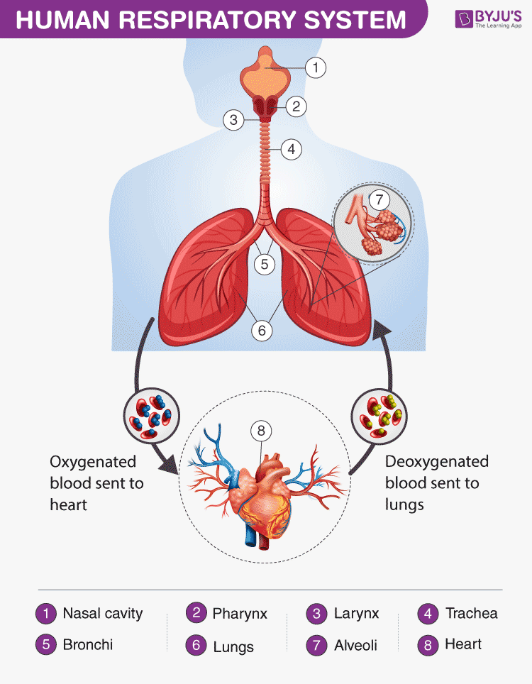
Human Respiratory System Diagram showing different parts of the Respiratory Tract
Features of the Human Respiratory System
The respiratory system in humans has the following important features:
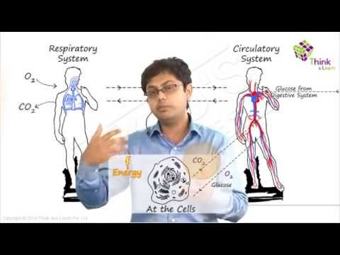
- The energy is generated by the breakdown of glucose molecules in all living cells of the human body.
- Oxygen is inhaled and is transported to various parts and are used in the process of burning food particles (breaking down glucose molecules) at the cellular level in a series of chemical reactions.
- The obtained glucose molecules are used for discharging energy in the form of ATP- (adenosine triphosphate)
Also Read: Difference between trachea and oesophagus
Respiratory System Parts and Functions
Let us have a detailed look at the different parts of the respiratory system and their functions.
Humans have exterior nostrils, which are divided by a framework of cartilaginous structure called the septum. This is the structure that separates the right nostril from the left nostril. Tiny hair follicles that cover the interior lining of nostrils act as the body’s first line of defence against foreign pathogens . Furthermore, they provide additional humidity for inhaled air.
Two cartilaginous chords lay the framework for the larynx. It is found in front of the neck and is responsible for vocals as well as aiding respiration. Hence, it is also informally called the voice box. When food is swallowed, a flap called the epiglottis folds over the top of the windpipe and prevents food from entering into the larynx.
Also check: What is the role of epiglottis and diaphragm in respiration?
The nasal chambers open up into a wide hollow space called the pharynx. It is a common passage for air as well as food. It functions by preventing the entry of food particles into the windpipe. The epiglottis is an elastic cartilage, which serves as a switch between the larynx and the oesophagus by allowing the passage of air into the lungs, and food in the gastrointestinal tract .
Have you ever wondered why we cough when we eat or swallow?
Talking while we eat or swallow may sometimes result in incessant coughing. The reason behind this reaction is the epiglottis. It is forced to open for the air to exit outwards and the food to enter into the windpipe, triggering a cough.
The trachea or the windpipe rises below the larynx and moves down to the neck. The walls of the trachea comprise C-shaped cartilaginous rings which give hardness to the trachea and maintain it by completely expanding. The trachea extends further down into the breastbone and splits into two bronchi, one for each lung.
The trachea splits into two tubes called the bronchi, which enter each lung individually. The bronchi divide into secondary and tertiary bronchioles, and it further branches out into small air-sacs called the alveoli. The alveoli are single-celled sacs of air with thin walls. It facilitates the exchange of oxygen and carbon dioxide molecules into or away from the bloodstream.
Lungs are the primary organs of respiration in humans and other vertebrates. They are located on either side of the heart, in the thoracic cavity of the chest. Anatomically, the lungs are spongy organs with an estimates total surface area between 50 to 75 sq meters. The primary function of the lungs is to facilitate the exchange of gases between the blood and the air. Interestingly, the right lung is quite bigger and heavier than the left lung.
Also Read: Respiration
The respiratory tract in humans is made up of the following parts:
- External nostrils – For the intake of air.
- Nasal chamber – which is lined with hair and mucus to filter the air from dust and dirt.
- Pharynx – It is a passage behind the nasal chamber and serves as the common passageway for both air and food.
- Larynx – Known as the soundbox as it houses the vocal chords, which are paramount in the generation of sound.
- Epiglottis – It is a flap-like structure that covers the glottis and prevents the entry of food into the windpipe.
- Trachea – It is a long tube passing through the mid-thoracic cavity.
- Bronchi – The trachea divides into left and right bronchi.
- Bronchioles – Each bronchus is further divided into finer channels known as bronchioles.
- Alveoli – The bronchioles terminate in balloon-like structures known as the alveoli.
- Lungs – Humans have a pair of lungs, which are sac-like structures and covered by a double-layered membrane known as pleura.
Air is inhaled with the help of nostrils, and in the nasal cavity, the air is cleansed by the fine hair follicles present within them. The cavity also has a group of blood vessels that warm the air. This air then passes to the pharynx, then to the larynx and into the trachea.
The trachea and the bronchi are coated with ciliated epithelial cells and goblet cells (secretory cells) which discharge mucus to moisten the air as it passes through the respiratory tract. It also traps the fine bits of dust or pathogen that escaped the hair in the nasal openings. The motile cilia beat in an ascending motion, such that the mucus and other foreign particles are carried back to the buccal cavity where it may either be coughed out (or swallowed.)
Once the air reaches the bronchus, it moves into the bronchioles, and then into the alveoli.
Respiratory System Functions
The functions of the human respiratory system are as follows:
Inhalation and Exhalation
The respiratory system helps in breathing (also known as pulmonary ventilation.) The air inhaled through the nose moves through the pharynx, larynx, trachea and into the lungs. The air is exhaled back through the same pathway. Changes in the volume and pressure in the lungs aid in pulmonary ventilation.
Exchange of Gases between Lungs and Bloodstream
Inside the lungs, the oxygen and carbon dioxide enter and exit respectively through millions of microscopic sacs called alveoli. The inhaled oxygen diffuses into the pulmonary capillaries, binds to haemoglobin and is pumped through the bloodstream. The carbon dioxide from the blood diffuses into the alveoli and is expelled through exhalation.
Also read: Exchange Of Gases in Plants
Exchange of Gases between Bloodstream and Body Tissues
The blood carries the oxygen from the lungs around the body and releases the oxygen when it reaches the capillaries. The oxygen is diffused through the capillary walls into the body tissues. The carbon dioxide also diffuses into the blood and is carried back to the lungs for release.
The Vibration of the Vocal Cords
While speaking, the muscles in the larynx move the arytenoid cartilage. These cartilages push the vocal cords together. During exhalation, when the air passes through the vocal cords, it makes them vibrate and creates sound.
Olfaction or Smelling
During inhalation, when the air enters the nasal cavities, some chemicals present in the air bind to it and activate the receptors of the nervous system on the cilia. The signals are sent to the olfactory bulbs via the brain.
Also Read: Respiratory System Disorders
Respiration is one of the metabolic processes which plays an essential role in all living organisms. However, lower organisms like the unicellular do not “breathe” like humans – intead, they utilise the process of diffusion. Annelids like earthworms have a moist cuticle which helps them in gaseous exchange. Respiration in fish occurs through special organs called gills. Most of the higher organisms possess a pair of lungs for breathing.
Also Read: Amphibolic Pathway
To learn more about respiration, check out the video below:

Frequently Asked Questions
What is the human respiratory system.
The human respiratory system is a system of organs responsible for inhaling oxygen and exhaling carbon dioxide in humans. The important respiratory organs in living beings include- lungs, gills, trachea, and skin.
What are the important respiratory system parts in humans?
The important human respiratory system parts include- Nose, larynx, pharynx, trachea, bronchi and lungs.
What is the respiratory tract made up of?
The respiratory tract is made up of nostrils, nasal chamber, larynx, pharynx, epiglottis, trachea, bronchioles, bronchi, alveoli, and lungs.

What are the main functions of the respiratory system?
The important functions of the respiratory system include- inhalation and exhalation of gases, exchange of gases between bloodstream and lungs, the gaseous exchange between bloodstream and body tissues, olfaction and vibration of vocal cords.
What are the different types of respiration in humans?
The different types of respiration in humans include- internal respiration, external respiration and cellular respiration. Internal respiration includes the exchange of gases between blood and cells, external respiration is the breathing process, whereas cellular respiration is the metabolic reactions taking place in the cells to produce energy.
What are the different stages of aerobic respiration?
Aerobic respiration is the process of breaking down glucose to produce energy. It occurs in the following different stages- glycolysis, pyruvate oxidation, citric acid cycle or Krebs cycle, and electron transport system.
Why do the cells need oxygen?
Our body cells require oxygen to release energy. The oxygen inhaled during respiration is used to break down the food to release energy.
What is the main difference between breathing and respiration in humans?
Breathing is the physical process of inhaling oxygen and exhaling carbon dioxide in and out of our lungs. On the contrary, respiration is the chemical process where oxygen is utilized to break down glucose to generate energy to carry out different cellular processes.
Explore more details about the human respiratory system or other related topics by registering at BYJU’S Biology

Put your understanding of this concept to test by answering a few MCQs. Click ‘Start Quiz’ to begin!
Select the correct answer and click on the “Finish” button Check your score and answers at the end of the quiz
Visit BYJU’S for all Biology related queries and study materials
Your result is as below
Request OTP on Voice Call
| BIOLOGY Related Links | |
Leave a Comment Cancel reply
Your Mobile number and Email id will not be published. Required fields are marked *
Post My Comment
Wow!!!!! Great notes . Thankyou byjus
Wow! this is really helpful. Thank you
Greet notes
Very helpful
Very nice information
good answers!
Thank you so much for the notes
Great notes helpful for students
The notes are really amazing. It helped me a lot. Thank you BYJU’S
The notes are amazing. It helped me a lot. Thank you Byju’s.
Thank you for the notes helpful in the revision
Very good notes. I love Byjus learning application
thank you byjus
These notes are very good for study, thank u so much Byjus.
Helpful for studying. Informative about the topic. Great notes!
Great job you are great Byjus and your notes is superb ❤❤❤❤❣❣❣❣❣🙏🙏🙏🙏
Register with BYJU'S & Download Free PDFs
Register with byju's & watch live videos.

An official website of the United States government
Here’s how you know
Official websites use .gov A .gov website belongs to an official government organization in the United States.
Secure .gov websites use HTTPS A lock ( A locked padlock ) or https:// means you’ve safely connected to the .gov website. Share sensitive information only on official, secure websites.
- Heart-Healthy Living
- High Blood Pressure
- Sickle Cell Disease
- Sleep Apnea
- Information & Resources on COVID-19
- The Heart Truth®
- Learn More Breathe Better®
- Blood Diseases and Disorders Education Program
- Publications and Resources
- Blood Disorders and Blood Safety
- Sleep Science and Sleep Disorders
- Lung Diseases
- Health Disparities and Inequities
- Heart and Vascular Diseases
- Precision Medicine Activities
- Obesity, Nutrition, and Physical Activity
- Population and Epidemiology Studies
- Women’s Health
- Research Topics
- Clinical Trials
- All Science A-Z
- Grants and Training Home
- Policies and Guidelines
- Funding Opportunities and Contacts
- Training and Career Development
- Email Alerts
- NHLBI in the Press
- Research Features
- Past Events
- Upcoming Events
- Mission and Strategic Vision
- Divisions, Offices and Centers
- Advisory Committees
- Budget and Legislative Information
- Jobs and Working at the NHLBI
- Contact and FAQs
- NIH Sleep Research Plan
- < Back To Health Topics
- How the Lungs Work
- The Respiratory System
- How Your Body Controls Breathing
- What Breathing Does for the Body
- How to Keep Your Lungs Healthy
MORE INFORMATION
How the Lungs Work The Lungs
Language switcher.
Your lungs are the pair of spongy, pinkish-gray organs in your chest.
When you inhale (breathe in), air enters your lungs, and oxygen from that air moves to your blood. At the same time, carbon dioxide, a waste gas, moves from your blood to the lungs and is exhaled (breathed out). This process, called gas exchange, is essential to life.
The lungs are the centerpiece of your respiratory system. Your respiratory system also includes the trachea (windpipe), muscles of the chest wall and diaphragm, blood vessels, and other tissues. All of these parts make breathing and gas exchange possible. Your brain controls your breathing rate (how fast or slow you breathe), by sensing your body’s need to get oxygen and also get rid of carbon dioxide.
Healthy lifestyle habits, such as physical activity and not smoking, can help prevent lung injury and disease.
Warning: The NCBI web site requires JavaScript to function. more...
An official website of the United States government
The .gov means it's official. Federal government websites often end in .gov or .mil. Before sharing sensitive information, make sure you're on a federal government site.
The site is secure. The https:// ensures that you are connecting to the official website and that any information you provide is encrypted and transmitted securely.
- Publications
- Account settings
- Browse Titles
NCBI Bookshelf. A service of the National Library of Medicine, National Institutes of Health.
StatPearls [Internet]. Treasure Island (FL): StatPearls Publishing; 2024 Jan-.

StatPearls [Internet].
Physiology, lung.
Moshe Haddad ; Sandeep Sharma .
Affiliations
Last Update: July 20, 2023 .
- Introduction
The lungs are the foundational organs of the respiratory system, whose most basic function is to facilitate gas exchange from the environment into the bloodstream. Oxygen gets transported through the alveoli into the capillary network, where it can enter the arterial system, ultimately perfuse tissue. The respiratory system is composed primarily of the nose, oropharynx, larynx, trachea, bronchi, bronchioles, and lungs. The lungs further divide into individual lobes, which ultimately subdivide into over 300 million alveoli. The alveoli are the primary location for gas exchange. The diaphragm is the primary respiratory muscle and receives innervation by the nerve roots of C3, C4, and C5 via the phrenic nerve. The external intercostals are inspiratory muscles used primarily during exercise and respiratory distress. The significant lung volumes/capacities and their definitions are listed below. [1] [2] [3] [4]
- Inspiratory reserve volume (IRV) : Volume that can be breathed after a normal inspiration
- Tidal volume (TV): Volume inspired and expired with each breath
- Expiratory reserve volume (ERV): Volume that can be expired after a normal breath
- Residual volume (RV) : Volume remaining in the lung after maximal expiration (cannot be measured by spirometry)
- Inspiratory capacity (IC) : Volume that can be breathed after normal exhalation
- Functional residual capacity (FRC) : Volume remaining in the lungs after normal expiration
- Vital capacity (VC) : Maximum volume able to be expired after maximal inspiration
- Total lung capacity (TLC) : Volume of air in the lungs after maximal inspiration
- Forced expiratory volume (FEV1) : Volume that can be expired in 1 second of maximum forced expiration
- Issues of Concern
The lung is a primary location for a large proportion of human diseases. Lung disease further classifies into obstructive and restrictive diseases.
Obstructive Disease
The definition of obstructive disease is lung disease with impaired expiration. It presents with decreased FVC, decreased FEV1, and, most notably, a dramatic decrease in FEV1/FVC. In obstructive disease, the air that should be expired is not, which leads to air trapping and an increased FRC. The two major examples of obstructive disease are listed below:
Asthma: a multifactorial disease characterized by chronic bronchial inflammation leading to eventual air trapping. Several key characteristics are as follows.
- Airway disease is mostly reversible (i.e., with the administration of a beta-agonist).
- Can cause chronic cough, wheezing, tachypnea, and dyspnea.
Chronic obstructive pulmonary disorder (COPD): a constellation of clinical symptoms that share features of both emphysema and chronic bronchitis leading to expiratory airflow limitation.
- Chronic bronchitis demonstrates long-term airway inflammation causing excessive cough and sputum production.
- Emphysema characteristically shows enlarged airspaces (loss of alveolar elasticity), leading to chronic dyspnea. The overly-distended air spaces prevent the lungs from adequately emptying.
- Smoking is the primary cause of the disease and is directly related to the severity of the disease course.
- Cigarettes induce inflammation in the lungs.
- Airways show small airway disease and parenchymal destruction.
Restrictive Disease
Restrictive lung disease is a lung disease in which restricted lung expansion causes decreased lung volumes. Its characteristics include both a decreased FVC and decreased FEV1; however, FVC is more reduced than FEV1 in restrictive disorders. So the ratio between them will increase. Several examples of restrictive lung disease are listed below [5] [1] [4] [6] [7] :
- Idiopathic pulmonary fibrosis
- Pneumoconiosis
- Sarcoidosis
- Cellular Level
Oxygen transport is the primary means by which the circulatory system perfuses tissue. Oxygen gets carried in the body in two major forms: bound to hemoglobin and dissolved. Hemoglobin is the major oxygen carrier in the body. The formula for the oxygen content of blood is as follows:
- CaO 2 = 1.34 x [Hgb] x (SaO 2 / 100) + 0.003 x PaO 2
- CaO 2 = oxygen content in the blood
- [Hb] = hemoglobin concentration
- SaO 2 = percentage of heme groups that are bound to oxygen
- PaO 2 = Partial pressure of oxygen
Four subunits comprise hemoglobin, each containing a heme-moiety that binds to iron. One molecule of O2 can bind to each iron atom of the heme group; therefore, each hemoglobin group can bind to four molecules of O 2 . [8] [9]
- Development
Lung development in-utero occurs in five main stages. The first stage begins with the development of the lung bud from the respiratory diverticulum during week 4 of embryogenesis. The stages are as follows [10] [11] [12] [13] :
- Embryonic : begins from weeks 4 to 7; this is when the formation of the major airways and pleura occur.
- Psuedoglandular : occurs during weeks 5 to 17; this is when the bronchial tree and respiratory parenchyma form.
- Canalicular : occurs during weeks 16 to 26; the distal airway, blood-air barrier, surfactant, and acini form.
- Saccular : occurs during weeks 24 to 38 weeks; the airspaces (alveoli) continue to expand.
- Alveolar : occurs from week 36 of gestation and throughout childhood. The alveoli become septated and more mature - improving airspace and capillary networks.
- Organ Systems Involved
The right ventricle conveys deoxygenated blood to the lungs via the pulmonary artery. The lungs will then breathe in air, which transfers oxygen through the alveoli into the dense capillary network surrounding the alveoli. The blood will then travel through the pulmonary vein into the left atrium, where it gets ejected from the left ventricle into the aorta. The aorta is the major artery that further branches into the system arteries, delivering blood to the cerebral, coronary, renal, splanchnic, and skin. Systemic veins return deoxygenated blood from these tissues to the vena cava, where it enters the right atrium and begins the cycle again.
The following equation can express blood flow dynamics: Q = P/R, where Q is the flow/cardiac output, P is the pressure gradient, and R is the total peripheral resistance. [14]
The function of the pulmonary system is to extract oxygen from the environment and provide it for aerobic respiration at the cellular level. Oxygen is ultimately used to produce ATP, and carbon dioxide is breathed out with other metabolic byproducts. [2]
Respiratory tract organs facilitate the process of gas exchange, including the nose, oral cavity, throat, trachea, bronchi, and lungs. The lungs divide into five major lobes: three lobes on the right and two lobes on the left. Each lobe is made up of many small alveoli, which are the primary site of gas exchange. At the alveoli, diffusion of gases into the arterioles occurs. [15]
The four significant aspects of respiratory mechanics are as follows: lung compliance, chest wall compliance, respiratory rate, and airway resistance. These work in conjunction to create a negative pressure within the lungs and pleural space, allowing air to be drawn into the lungs. Conversely, drops in lung volume increase pressure in the lungs, which forces air out.
The following equation represents lung compliance: C = V/P, where C is lung compliance, V is lung volume, and P is lung pressure.
The respiratory rate will adjust to meet the needs of the body. As oxygen demand increases and carbon dioxide builds up (i.e., exercise), the lungs will accommodate by increasing the respiratory rate to facilitate the diffusion of these gases.
Chest wall compliance counteracts the force of lung compliance, attempting to increase lung volume (contrary to lung compliance, where the lungs attempt to decrease volume)
Poiseuille’s law describes airway resistance, the law states: R = (8*n*l)/(pi * r ^4), where R = resistance, n = viscosity of air, l = length of the airway, and r = radius of the airway. It is essential to note the inverse fourth power relationship between the radius of the airway and the resistance. Small adjustments in airway radius will substantially alter the resistance. [2]
- Related Testing
Pulmonary function tests (PFTs) serve the purpose of both the investigation and monitoring of patients with pulmonary disease. They provide information relating to both large and small airways, lung parenchyma, and pulmonary capillary bed. PFTs are a safe and effective way to help the clinician diagnose and monitor pathology. There are many indications for PFTs, including:
- Chest X-ray abnormalities
- Shortness of breath
- Monitoring of patients with known respiratory disease (ie, COPD, asthma, interstitial fibrosis)
- Monitoring of diseases that may have associated respiratory complications (ie, Guillain-Barré)
- Perioperative evaluation
- Lung transplant surveillance
The major measurements included in pulmonary function testing include FEV1, FVC, and FEV1/FVC. Some of the more common findings on PFTs are as follows. [16]
Interstitial fibrosis: dec. FVC, dec. FEV1, normal/ inc. FEV1/FVC
Asthma: dec. FVC, dec. FEV1, dec. FEV1/FVC
COPD: dec. FVC, dec. FEV1, dec. FEV1/FVC
- Pathophysiology
COPD is one of the major leading causes of lung pathology worldwide. Tobacco smoke is the primary cause of the disease. Nicotine and the thousands of other toxic components contained in cigarettes lead to the formation of free radicals, causing alveolar damage.
COPD is a general term referring to the combination of chronic bronchitis and emphysema. It is an obstructive disease, impairing expiration. The chronic bronchitis component causes severe hypoxemia due to airway inflammation and mucus production. The emphysematous component causes enlargement of air spaces, leading to decreased recoil and increased compliance, which ultimately causes air trapping. [17] [18]
- Clinical Significance
As lung pathology remains a significant cause of morbidity and mortality nationwide, understanding of basic lung physiology and its ties to pathology is essential for the clinician. Dysfunction of the pulmonary system ultimately leads to hypoxia. There are four classifications of hypoxia etiology: hypoventilation, right-to-left-shunt, V/Q mismatch, and diffusion limitations.
Hypoventilation:
Any disease that decreases the ventilatory rate of alveoli ultimately results in hypoventilation. Several causes include obesity, CNS depressants, rib fractures, neurologic defects, and kyphoscoliosis.
Right-to-left-shunt:
The right-to-left shunt is when deoxygenated blood bypasses the lungs from the right heart to the left heart. This situation can occur both anatomically and physiologically. Anatomical causes of anatomical shunts include congenital heart malformations (i.e., tetralogy of Fallot) and arteriovenous malformations. Physiologic shunts occur when there is blood flow to an unventilated alveolus; this occurs in instances where air cannot diffuse across the alveolus, such as acute respiratory distress syndrome or pleural effusions.
V/Q mismatches:
V/Q mismatches occur when blood flow and ventilation are mismatched, as the name implies. Ideally, ventilation and perfusion match for proper gas exchange. The normal V/Q ratio is 0.8. A high V/Q ratio occurs when ventilation exceeds perfusion. The classic example of this would be in a pulmonary embolism, where perfusion is completely blocked to a segment of the lung while ventilation remains unaffected. A low V/Q ratio occurs when there is normal blood flow to a region of the lung, but ventilation is blocked. An example of this would be in COPD, where the alveolus’ surface area is decreased, causing inadequate gas exchange.
Diffusion limitations:
Diffusion limitation occurs when oxygen cannot effectively move from the alveoli into the pulmonary capillaries. This condition can take place when there is the destruction of the alveoli (i.e., COPD), or in instances where there is destruction or thickening of the lung parenchyma (i.e., interstitial fibrosis). These commonly occur in conjunction with V/Q mismatches. [19] [2]
- Review Questions
- Access free multiple choice questions on this topic.
- Comment on this article.
Disclosure: Moshe Haddad declares no relevant financial relationships with ineligible companies.
Disclosure: Sandeep Sharma declares no relevant financial relationships with ineligible companies.
This book is distributed under the terms of the Creative Commons Attribution-NonCommercial-NoDerivatives 4.0 International (CC BY-NC-ND 4.0) ( http://creativecommons.org/licenses/by-nc-nd/4.0/ ), which permits others to distribute the work, provided that the article is not altered or used commercially. You are not required to obtain permission to distribute this article, provided that you credit the author and journal.
- Cite this Page Haddad M, Sharma S. Physiology, Lung. [Updated 2023 Jul 20]. In: StatPearls [Internet]. Treasure Island (FL): StatPearls Publishing; 2024 Jan-.
In this Page
Bulk download.
- Bulk download StatPearls data from FTP
Related information
- PMC PubMed Central citations
- PubMed Links to PubMed
Similar articles in PubMed
- Physiology, Functional Residual Capacity. [StatPearls. 2024] Physiology, Functional Residual Capacity. Hopkins E, Sharma S. StatPearls. 2024 Jan
- Physiology, Lung Capacity. [StatPearls. 2024] Physiology, Lung Capacity. Delgado BJ, Bajaj T. StatPearls. 2024 Jan
- Vital Capacity. [StatPearls. 2024] Vital Capacity. David S, Sharma S. StatPearls. 2024 Jan
- Review [Physiopathology of acute respiratory failure in COPD and asthma]. [Minerva Anestesiol. 2001] Review [Physiopathology of acute respiratory failure in COPD and asthma]. Mergoni M, Rossi A. Minerva Anestesiol. 2001 Apr; 67(4):198-205.
- Review Medical devices in allergy practice. [World Allergy Organ J. 2020] Review Medical devices in allergy practice. Popov TA, Passalacqua G, González-Díaz SN, Plavec D, Braido F, García-Abujeta JL, Dubuske L, Rouadi P, Morais-Almeida M, Bonini S, et al. World Allergy Organ J. 2020 Oct; 13(10):100466. Epub 2020 Sep 30.
Recent Activity
- Physiology, Lung - StatPearls Physiology, Lung - StatPearls
Your browsing activity is empty.
Activity recording is turned off.
Turn recording back on
Connect with NLM
National Library of Medicine 8600 Rockville Pike Bethesda, MD 20894
Web Policies FOIA HHS Vulnerability Disclosure
Help Accessibility Careers

- school Campus Bookshelves
- menu_book Bookshelves
- perm_media Learning Objects
- login Login
- how_to_reg Request Instructor Account
- hub Instructor Commons
Margin Size
- Download Page (PDF)
- Download Full Book (PDF)
- Periodic Table
- Physics Constants
- Scientific Calculator
- Reference & Cite
- Tools expand_more
- Readability
selected template will load here
This action is not available.

16.3: Circulatory and Respiratory Systems
- Last updated
- Save as PDF
- Page ID 7073

\( \newcommand{\vecs}[1]{\overset { \scriptstyle \rightharpoonup} {\mathbf{#1}} } \)
\( \newcommand{\vecd}[1]{\overset{-\!-\!\rightharpoonup}{\vphantom{a}\smash {#1}}} \)
\( \newcommand{\id}{\mathrm{id}}\) \( \newcommand{\Span}{\mathrm{span}}\)
( \newcommand{\kernel}{\mathrm{null}\,}\) \( \newcommand{\range}{\mathrm{range}\,}\)
\( \newcommand{\RealPart}{\mathrm{Re}}\) \( \newcommand{\ImaginaryPart}{\mathrm{Im}}\)
\( \newcommand{\Argument}{\mathrm{Arg}}\) \( \newcommand{\norm}[1]{\| #1 \|}\)
\( \newcommand{\inner}[2]{\langle #1, #2 \rangle}\)
\( \newcommand{\Span}{\mathrm{span}}\)
\( \newcommand{\id}{\mathrm{id}}\)
\( \newcommand{\kernel}{\mathrm{null}\,}\)
\( \newcommand{\range}{\mathrm{range}\,}\)
\( \newcommand{\RealPart}{\mathrm{Re}}\)
\( \newcommand{\ImaginaryPart}{\mathrm{Im}}\)
\( \newcommand{\Argument}{\mathrm{Arg}}\)
\( \newcommand{\norm}[1]{\| #1 \|}\)
\( \newcommand{\Span}{\mathrm{span}}\) \( \newcommand{\AA}{\unicode[.8,0]{x212B}}\)
\( \newcommand{\vectorA}[1]{\vec{#1}} % arrow\)
\( \newcommand{\vectorAt}[1]{\vec{\text{#1}}} % arrow\)
\( \newcommand{\vectorB}[1]{\overset { \scriptstyle \rightharpoonup} {\mathbf{#1}} } \)
\( \newcommand{\vectorC}[1]{\textbf{#1}} \)
\( \newcommand{\vectorD}[1]{\overrightarrow{#1}} \)
\( \newcommand{\vectorDt}[1]{\overrightarrow{\text{#1}}} \)
\( \newcommand{\vectE}[1]{\overset{-\!-\!\rightharpoonup}{\vphantom{a}\smash{\mathbf {#1}}}} \)
Animals are complex multicellular organisms that require a mechanism for transporting nutrients throughout their bodies and removing wastes. The human circulatory system has a complex network of blood vessels that reach all parts of the body. This extensive network supplies the cells, tissues, and organs with oxygen and nutrients, and removes carbon dioxide and waste compounds.
The medium for transport of gases and other molecules is the blood, which continually circulates through the system. Pressure differences within the system cause the movement of the blood and are created by the pumping of the heart.
Gas exchange between tissues and the blood is an essential function of the circulatory system. In humans, other mammals, and birds, blood absorbs oxygen and releases carbon dioxide in the lungs. Thus the circulatory and respiratory system, whose function is to obtain oxygen and discharge carbon dioxide, work in tandem.
The Respiratory System
Take a breath in and hold it. Wait several seconds and then let it out. Humans, when they are not exerting themselves, breathe approximately 15 times per minute on average. This equates to about 900 breaths an hour or 21,600 breaths per day. With every inhalation, air fills the lungs, and with every exhalation, it rushes back out. That air is doing more than just inflating and deflating the lungs in the chest cavity. The air contains oxygen that crosses the lung tissue, enters the bloodstream, and travels to organs and tissues. There, oxygen is exchanged for carbon dioxide, which is a cellular waste material. Carbon dioxide exits the cells, enters the bloodstream, travels back to the lungs, and is expired out of the body during exhalation.
Breathing is both a voluntary and an involuntary event. How often a breath is taken and how much air is inhaled or exhaled is regulated by the respiratory center in the brain in response to signals it receives about the carbon dioxide content of the blood. However, it is possible to override this automatic regulation for activities such as speaking, singing and swimming under water.
During inhalation the diaphragm descends creating a negative pressure around the lungs and they begin to inflate, drawing in air from outside the body. The air enters the body through the nasal cavity located just inside the nose (Figure \(\PageIndex{1}\)). As the air passes through the nasal cavity, the air is warmed to body temperature and humidified by moisture from mucous membranes. These processes help equilibrate the air to the body conditions, reducing any damage that cold, dry air can cause. Particulate matter that is floating in the air is removed in the nasal passages by hairs, mucus, and cilia. Air is also chemically sampled by the sense of smell.
From the nasal cavity, air passes through the pharynx (throat) and the larynx (voice box) as it makes its way to the trachea (Figure \(\PageIndex{1}\)). The main function of the trachea is to funnel the inhaled air to the lungs and the exhaled air back out of the body. The human trachea is a cylinder, about 25 to 30 cm (9.8–11.8 in) long, which sits in front of the esophagus and extends from the pharynx into the chest cavity to the lungs. It is made of incomplete rings of cartilage and smooth muscle. The cartilage provides strength and support to the trachea to keep the passage open. The trachea is lined with cells that have cilia and secrete mucus. The mucus catches particles that have been inhaled, and the cilia move the particles toward the pharynx.
The end of the trachea divides into two bronchi that enter the right and left lung. Air enters the lungs through the primary bronchi. The primary bronchus divides, creating smaller and smaller diameter bronchi until the passages are under 1 mm (.03 in) in diameter when they are called bronchioles as they split and spread through the lung. Like the trachea, the bronchus and bronchioles are made of cartilage and smooth muscle. Bronchi are innervated by nerves of both the parasympathetic and sympathetic nervous systems that control muscle contraction (parasympathetic) or relaxation (sympathetic) in the bronchi and bronchioles, depending on the nervous system’s cues. The final bronchioles are the respiratory bronchioles. Alveolar ducts are attached to the end of each respiratory bronchiole. At the end of each duct are alveolar sacs, each containing 20 to 30 alveoli. Gas exchange occurs only in the alveoli. The alveoli are thin-walled and look like tiny bubbles within the sacs. The alveoli are in direct contact with capillaries of the circulatory system. Such intimate contact ensures that oxygen will diffuse from the alveoli into the blood. In addition, carbon dioxide will diffuse from the blood into the alveoli to be exhaled. The anatomical arrangement of capillaries and alveoli emphasizes the structural and functional relationship of the respiratory and circulatory systems. Estimates for the surface area of alveoli in the lungs vary around 100 m 2 . This large area is about the area of half a tennis court. This large surface area, combined with the thin-walled nature of the alveolar cells, allows gases to easily diffuse across the cells.
ART CONNECTION
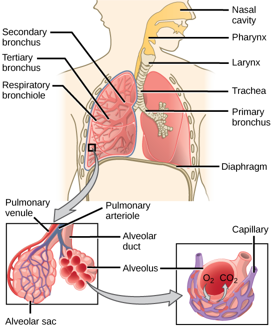
Which of the following statements about the human respiratory system is false?
- When we breathe in, air travels from the pharynx to the trachea.
- The bronchioles branch into bronchi.
- Alveolar ducts connect to alveolar sacs.
- Gas exchange between the lungs and blood takes place in the alveolus.
CONCEPT IN ACTION
Watch this video for a review of the respiratory system.
The Circulatory System
The circulatory system is a network of vessels—the arteries, veins, and capillaries—and a pump, the heart. In all vertebrate organisms this is a closed-loop system, in which the blood is largely separated from the body’s other extracellular fluid compartment, the interstitial fluid, which is the fluid bathing the cells. Blood circulates inside blood vessels and circulates unidirectionally from the heart around one of two circulatory routes, then returns to the heart again; this is a closed circulatory system. Open circulatory systems are found in invertebrate animals in which the circulatory fluid bathes the internal organs directly even though it may be moved about with a pumping heart.
The heart is a complex muscle that consists of two pumps: one that pumps blood through pulmonary circulation to the lungs, and the other that pumps blood through systemic circulation to the rest of the body’s tissues (and the heart itself).
The heart is asymmetrical, with the left side being larger than the right side, correlating with the different sizes of the pulmonary and systemic circuits (Figure \(\PageIndex{2}\)). In humans, the heart is about the size of a clenched fist; it is divided into four chambers: two atria and two ventricles. There is one atrium and one ventricle on the right side and one atrium and one ventricle on the left side. The right atrium receives deoxygenated blood from the systemic circulation through the major veins: the superior vena cava, which drains blood from the head and from the veins that come from the arms, as well as the inferior vena cava, which drains blood from the veins that come from the lower organs and the legs. This deoxygenated blood then passes to the right ventricle through the tricuspid valve, which prevents the backflow of blood. After it is filled, the right ventricle contracts, pumping the blood to the lungs for reoxygenation. The left atrium receives the oxygen-rich blood from the lungs. This blood passes through the bicuspid valve to the left ventricle where the blood is pumped into the aorta. The aorta is the major artery of the body, taking oxygenated blood to the organs and muscles of the body. This pattern of pumping is referred to as double circulation and is found in all mammals. (Figure \(\PageIndex{2}\)).
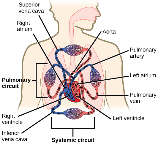
Which of the following statements about the circulatory system is false?
- Blood in the pulmonary vein is deoxygenated.
- Blood in the inferior vena cava is deoxygenated.
- Blood in the pulmonary artery is deoxygenated.
- Blood in the aorta is oxygenated.
The Cardiac Cycle
The main purpose of the heart is to pump blood through the body; it does so in a repeating sequence called the cardiac cycle. The cardiac cycle is the flow of blood through the heart coordinated by electrochemical signals that cause the heart muscle to contract and relax. In each cardiac cycle, a sequence of contractions pushes out the blood, pumping it through the body; this is followed by a relaxation phase, where the heart fills with blood. These two phases are called the systole (contraction) and diastole (relaxation), respectively (Figure \(\PageIndex{3}\)). The signal for contraction begins at a location on the outside of the right atrium. The electrochemical signal moves from there across the atria causing them to contract. The contraction of the atria forces blood through the valves into the ventricles. Closing of these valves caused by the contraction of the ventricles produces a “lub” sound. The signal has, by this time, passed down the walls of the heart, through a point between the right atrium and right ventricle. The signal then causes the ventricles to contract. The ventricles contract together forcing blood into the aorta and the pulmonary arteries. Closing of the valves to these arteries caused by blood being drawn back toward the heart during ventricular relaxation produces a monosyllabic “dub” sound.
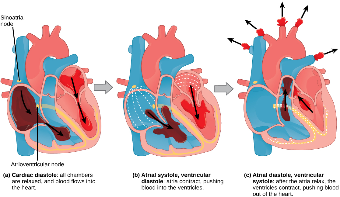
The pumping of the heart is a function of the cardiac muscle cells, or cardiomyocytes, that make up the heart muscle. Cardiomyocytes are distinctive muscle cells that are striated like skeletal muscle but pump rhythmically and involuntarily like smooth muscle; adjacent cells are connected by intercalated disks found only in cardiac muscle. These connections allow the electrical signal to travel directly to neighboring muscle cells.
The electrical impulses in the heart produce electrical currents that flow through the body and can be measured on the skin using electrodes. This information can be observed as an electrocardiogram (ECG) a recording of the electrical impulses of the cardiac muscle.

Visit the following website to see the heart’s pacemaker, or electrocardiogram system, in action.
Blood Vessels
The blood from the heart is carried through the body by a complex network of blood vessels (Figure \(\PageIndex{4}\)). Arteries take blood away from the heart. The main artery of the systemic circulation is the aorta; it branches into major arteries that take blood to different limbs and organs. The aorta and arteries near the heart have heavy but elastic walls that respond to and smooth out the pressure differences caused by the beating heart. Arteries farther away from the heart have more muscle tissue in their walls that can constrict to affect flow rates of blood. The major arteries diverge into minor arteries, and then smaller vessels called arterioles, to reach more deeply into the muscles and organs of the body.
Arterioles diverge into capillary beds. Capillary beds contain a large number, 10’s to 100’s of capillaries that branch among the cells of the body. Capillaries are narrow-diameter tubes that can fit single red blood cells and are the sites for the exchange of nutrients, waste, and oxygen with tissues at the cellular level. Fluid also leaks from the blood into the interstitial space from the capillaries. The capillaries converge again into venules that connect to minor veins that finally connect to major veins. Veins are blood vessels that bring blood high in carbon dioxide back to the heart. Veins are not as thick-walled as arteries, since pressure is lower, and they have valves along their length that prevent backflow of blood away from the heart. The major veins drain blood from the same organs and limbs that the major arteries supply.
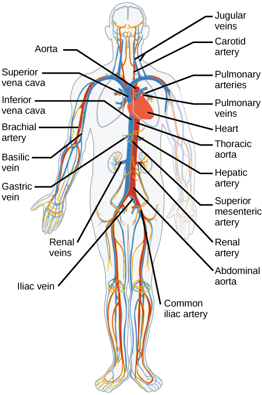
Section Summary
Animal respiratory systems are designed to facilitate gas exchange. In mammals, air is warmed and humidified in the nasal cavity. Air then travels down the pharynx and larynx, through the trachea, and into the lungs. In the lungs, air passes through the branching bronchi, reaching the respiratory bronchioles. The respiratory bronchioles open up into the alveolar ducts, alveolar sacs, and alveoli. Because there are so many alveoli and alveolar sacs in the lung, the surface area for gas exchange is very large.
The mammalian circulatory system is a closed system with double circulation passing through the lungs and the body. It consists of a network of vessels containing blood that circulates because of pressure differences generated by the heart.
The heart contains two pumps that move blood through the pulmonary and systemic circulations. There is one atrium and one ventricle on the right side and one atrium and one ventricle on the left side. The pumping of the heart is a function of cardiomyocytes, distinctive muscle cells that are striated like skeletal muscle but pump rhythmically and involuntarily like smooth muscle. The signal for contraction begins in the wall of the right atrium. The electrochemical signal causes the two atria to contract in unison; then the signal causes the ventricles to contract. The blood from the heart is carried through the body by a complex network of blood vessels; arteries take blood away from the heart, and veins bring blood back to the heart.
Art Connections
Figure \(\PageIndex{1}\): Which of the following statements about the human respiratory system is false?
A. When we breathe in, air travels from the pharynx to the trachea. B. The bronchioles branch into bronchi. C. Alveolar ducts connect to alveolar sacs. D. Gas exchange between the lungs and blood takes place in the alveolus.
Figure \(\PageIndex{2}\): Which of the following statements about the circulatory system is false?
A. Blood in the pulmonary vein is deoxygenated. B. Blood in the inferior vena cava is deoxygenated. C. Blood in the pulmonary artery is deoxygenated. D. Blood in the aorta is oxygenated.
Contributors and Attributions
Samantha Fowler (Clayton State University), Rebecca Roush (Sandhills Community College), James Wise (Hampton University). Original content by OpenStax (CC BY 4.0; Access for free at https://cnx.org/contents/b3c1e1d2-83...4-e119a8aafbdd ).
Critical Thinking Questions
Describe the three regions of the pharynx and their functions.
If a person sustains an injury to the epiglottis, what would be the physiological result?
Compare and contrast the conducting and respiratory zones.
Compare and contrast the right and left lungs.
Why are the pleurae not damaged during normal breathing?
Describe what is meant by the term “lung compliance.”
Outline the steps involved in quiet breathing.
What is respiratory rate and how is it controlled?
Compare and contrast Dalton’s law and Henry’s law.
A smoker develops damage to several alveoli that then can no longer function. How does this affect gas exchange?
Compare and contrast adult hemoglobin and fetal hemoglobin.
Describe the relationship between the partial pressure of oxygen and the binding of oxygen to hemoglobin.
Describe three ways in which carbon dioxide can be transported.
Describe the neural factors involved in increasing ventilation during exercise.
What is the major mechanism that results in acclimatization?
During what timeframe does a fetus have enough mature structures to breathe on its own if born prematurely? Describe the other structures that develop during this phase.
Describe fetal breathing movements and their purpose.
As an Amazon Associate we earn from qualifying purchases.
This book may not be used in the training of large language models or otherwise be ingested into large language models or generative AI offerings without OpenStax's permission.
Want to cite, share, or modify this book? This book uses the Creative Commons Attribution License and you must attribute OpenStax.
Access for free at https://openstax.org/books/anatomy-and-physiology/pages/1-introduction
- Authors: J. Gordon Betts, Kelly A. Young, James A. Wise, Eddie Johnson, Brandon Poe, Dean H. Kruse, Oksana Korol, Jody E. Johnson, Mark Womble, Peter DeSaix
- Publisher/website: OpenStax
- Book title: Anatomy and Physiology
- Publication date: Apr 25, 2013
- Location: Houston, Texas
- Book URL: https://openstax.org/books/anatomy-and-physiology/pages/1-introduction
- Section URL: https://openstax.org/books/anatomy-and-physiology/pages/22-critical-thinking-questions
© Jan 27, 2022 OpenStax. Textbook content produced by OpenStax is licensed under a Creative Commons Attribution License . The OpenStax name, OpenStax logo, OpenStax book covers, OpenStax CNX name, and OpenStax CNX logo are not subject to the Creative Commons license and may not be reproduced without the prior and express written consent of Rice University.
COVID-19: Long-term effects
Some people continue to experience health problems long after having COVID-19. Understand the possible symptoms and risk factors for post-COVID-19 syndrome.
Most people who get coronavirus disease 2019 (COVID-19) recover within a few weeks. But some people — even those who had mild versions of the disease — might have symptoms that last a long time afterward. These ongoing health problems are sometimes called post- COVID-19 syndrome, post- COVID conditions, long COVID-19 , long-haul COVID-19 , and post acute sequelae of SARS COV-2 infection (PASC).
What is post-COVID-19 syndrome and how common is it?
Post- COVID-19 syndrome involves a variety of new, returning or ongoing symptoms that people experience more than four weeks after getting COVID-19 . In some people, post- COVID-19 syndrome lasts months or years or causes disability.
Research suggests that between one month and one year after having COVID-19 , 1 in 5 people ages 18 to 64 has at least one medical condition that might be due to COVID-19 . Among people age 65 and older, 1 in 4 has at least one medical condition that might be due to COVID-19 .
What are the symptoms of post-COVID-19 syndrome?
The most commonly reported symptoms of post- COVID-19 syndrome include:
- Symptoms that get worse after physical or mental effort
- Lung (respiratory) symptoms, including difficulty breathing or shortness of breath and cough
Other possible symptoms include:
- Neurological symptoms or mental health conditions, including difficulty thinking or concentrating, headache, sleep problems, dizziness when you stand, pins-and-needles feeling, loss of smell or taste, and depression or anxiety
- Joint or muscle pain
- Heart symptoms or conditions, including chest pain and fast or pounding heartbeat
- Digestive symptoms, including diarrhea and stomach pain
- Blood clots and blood vessel (vascular) issues, including a blood clot that travels to the lungs from deep veins in the legs and blocks blood flow to the lungs (pulmonary embolism)
- Other symptoms, such as a rash and changes in the menstrual cycle
Keep in mind that it can be hard to tell if you are having symptoms due to COVID-19 or another cause, such as a preexisting medical condition.
It's also not clear if post- COVID-19 syndrome is new and unique to COVID-19 . Some symptoms are similar to those caused by chronic fatigue syndrome and other chronic illnesses that develop after infections. Chronic fatigue syndrome involves extreme fatigue that worsens with physical or mental activity, but doesn't improve with rest.
Why does COVID-19 cause ongoing health problems?
Organ damage could play a role. People who had severe illness with COVID-19 might experience organ damage affecting the heart, kidneys, skin and brain. Inflammation and problems with the immune system can also happen. It isn't clear how long these effects might last. The effects also could lead to the development of new conditions, such as diabetes or a heart or nervous system condition.
The experience of having severe COVID-19 might be another factor. People with severe symptoms of COVID-19 often need to be treated in a hospital intensive care unit. This can result in extreme weakness and post-traumatic stress disorder, a mental health condition triggered by a terrifying event.
What are the risk factors for post-COVID-19 syndrome?
You might be more likely to have post- COVID-19 syndrome if:
- You had severe illness with COVID-19 , especially if you were hospitalized or needed intensive care.
- You had certain medical conditions before getting the COVID-19 virus.
- You had a condition affecting your organs and tissues (multisystem inflammatory syndrome) while sick with COVID-19 or afterward.
Post- COVID-19 syndrome also appears to be more common in adults than in children and teens. However, anyone who gets COVID-19 can have long-term effects, including people with no symptoms or mild illness with COVID-19 .
What should you do if you have post-COVID-19 syndrome symptoms?
If you're having symptoms of post- COVID-19 syndrome, talk to your health care provider. To prepare for your appointment, write down:
- When your symptoms started
- What makes your symptoms worse
- How often you experience symptoms
- How your symptoms affect your activities
Your health care provider might do lab tests, such as a complete blood count or liver function test. You might have other tests or procedures, such as chest X-rays, based on your symptoms. The information you provide and any test results will help your health care provider come up with a treatment plan.
In addition, you might benefit from connecting with others in a support group and sharing resources.
- Long COVID or post-COVID conditions. Centers for Disease Control and Prevention. https://www.cdc.gov/coronavirus/2019-ncov/long-term-effects.html. Accessed May 6, 2022.
- Post-COVID conditions: Overview for healthcare providers. Centers for Disease Control and Prevention. https://www.cdc.gov/coronavirus/2019-ncov/hcp/clinical-care/post-covid-conditions.html. Accessed May 6, 2022.
- Mikkelsen ME, et al. COVID-19: Evaluation and management of adults following acute viral illness. https://www.uptodate.com/contents/search. Accessed May 6, 2022.
- Saeed S, et al. Coronavirus disease 2019 and cardiovascular complications: Focused clinical review. Journal of Hypertension. 2021; doi:10.1097/HJH.0000000000002819.
- AskMayoExpert. Post-COVID-19 syndrome. Mayo Clinic; 2022.
- Multisystem inflammatory syndrome (MIS). Centers for Disease Control and Prevention. https://www.cdc.gov/mis/index.html. Accessed May 24, 2022.
- Patient tips: Healthcare provider appointments for post-COVID conditions. https://www.cdc.gov/coronavirus/2019-ncov/long-term-effects/post-covid-appointment/index.html. Accessed May 24, 2022.
- Bull-Otterson L, et al. Post-COVID conditions among adult COVID-19 survivors aged 18-64 and ≥ 65 years — United States, March 2020 — November 2021. MMWR Morbidity and Mortality Weekly Report. 2022; doi:10.15585/mmwr.mm7121e1.
Products and Services
- A Book: Endemic - A Post-Pandemic Playbook
- Begin Exploring Women's Health Solutions at Mayo Clinic Store
- A Book: Future Care
- Antibiotics: Are you misusing them?
- COVID-19 and vitamin D
- Convalescent plasma therapy
- Coronavirus disease 2019 (COVID-19)
- COVID-19: How can I protect myself?
- Herd immunity and respiratory illness
- COVID-19 and pets
- COVID-19 and your mental health
- COVID-19 antibody testing
- COVID-19, cold, allergies and the flu
- COVID-19 tests
- COVID-19 drugs: Are there any that work?
- COVID-19 in babies and children
- Coronavirus infection by race
- COVID-19 travel advice
- COVID-19 vaccine: Should I reschedule my mammogram?
- COVID-19 vaccines for kids: What you need to know
- COVID-19 vaccines
- COVID-19 variant
- COVID-19 vs. flu: Similarities and differences
- COVID-19: Who's at higher risk of serious symptoms?
- Debunking coronavirus myths
- Different COVID-19 vaccines
- Extracorporeal membrane oxygenation (ECMO)
- Fever: First aid
- Fever treatment: Quick guide to treating a fever
- Fight coronavirus (COVID-19) transmission at home
- Honey: An effective cough remedy?
- How do COVID-19 antibody tests differ from diagnostic tests?
- How to measure your respiratory rate
- How to take your pulse
- How to take your temperature
- How well do face masks protect against COVID-19?
- Is hydroxychloroquine a treatment for COVID-19?
- Loss of smell
- Mayo Clinic Minute: You're washing your hands all wrong
- Mayo Clinic Minute: How dirty are common surfaces?
- Multisystem inflammatory syndrome in children (MIS-C)
- Nausea and vomiting
- Pregnancy and COVID-19
- Safe outdoor activities during the COVID-19 pandemic
- Safety tips for attending school during COVID-19
- Sex and COVID-19
- Shortness of breath
- Thermometers: Understand the options
- Treating COVID-19 at home
- Unusual symptoms of coronavirus
- Vaccine guidance from Mayo Clinic
- Watery eyes
Related information
- Post-COVID Recovery & COVID-19 Support Group - Related information Post-COVID Recovery & COVID-19 Support Group
- Rehabilitation after COVID-19 - Related information Rehabilitation after COVID-19
- Post-COVID-19 syndrome could be a long haul (podcast) - Related information Post-COVID-19 syndrome could be a long haul (podcast)
- COVID-19 Coronavirus Long-term effects
We’re transforming healthcare
Make a gift now and help create new and better solutions for more than 1.3 million patients who turn to Mayo Clinic each year.
- Share full article
Advertisement
Supported by
Guest Essay
Why the New Human Case of Bird Flu Is So Alarming

By Rick Bright
Dr. Bright is a virologist and the former head of the Biomedical Advanced Research and Development Authority.
The third human case of H5N1, reported on Thursday in a farmworker in Michigan who was experiencing respiratory symptoms, tells us that the current bird flu situation is at a dangerous inflection point.
The virus is adapting in predictable ways that increase its risk to humans, reflecting our failure to contain it early on. The solutions to this brewing crisis — such as comprehensive testing — have been there all along, and they’re becoming only more important. If we keep ignoring the warning signs we have only ourselves to blame.
H5N1 has long been more than a bird problem. The virus has found its way into dairy cattle across nine states , affecting 69 herds that we know about. Of the three human cases of H5N1 that have been identified, all involve farmworkers who were in direct contact with infected cows or milk. The first two cases were relatively mild, involving symptoms like eye irritation, or conjunctivitis. However, the most recent case has shown more concerning signs, including coughing.
The emergence of respiratory symptoms is disconcerting because it indicates a potential shift in how the virus affects humans. Coughing can spread viruses more easily than eye irritation can.
New symptoms should be expected as the virus continues to spread and adapt to humans. Yet our response to this looming danger has been woefully inadequate, particularly in the area of testing.
Testing is our first line of defense in identifying and controlling infectious diseases. It allows health responders to understand the extent of an outbreak, identify who is infected and take measures to prevent further spread.
We are having trouble retrieving the article content.
Please enable JavaScript in your browser settings.
Thank you for your patience while we verify access. If you are in Reader mode please exit and log into your Times account, or subscribe for all of The Times.
Thank you for your patience while we verify access.
Already a subscriber? Log in .
Want all of The Times? Subscribe .
Official websites use .gov
A .gov website belongs to an official government organization in the United States.
Secure .gov websites use HTTPS
A lock ( ) or https:// means you've safely connected to the .gov website. Share sensitive information only on official, secure websites.
CDC updates and simplifies respiratory virus recommendations
Recommendations are easier to follow and help protect those most at risk
For Immediate Release: Friday, March 1, 2024 Contact: Media Relations (404) 639-3286
CDC released today updated recommendations for how people can protect themselves and their communities from respiratory viruses, including COVID-19. The new guidance brings a unified approach to addressing risks from a range of common respiratory viral illnesses, such as COVID-19, flu, and RSV, which can cause significant health impacts and strain on hospitals and health care workers. CDC is making updates to the recommendations now because the U.S. is seeing far fewer hospitalizations and deaths associated with COVID-19 and because we have more tools than ever to combat flu, COVID, and RSV.
“Today’s announcement reflects the progress we have made in protecting against severe illness from COVID-19,” said CDC Director Dr. Mandy Cohen. “However, we still must use the commonsense solutions we know work to protect ourselves and others from serious illness from respiratory viruses—this includes vaccination, treatment, and staying home when we get sick.”
As part of the guidance, CDC provides active recommendations on core prevention steps and strategies:
- Staying up to date with vaccination to protect people against serious illness, hospitalization, and death. This includes flu, COVID-19, and RSV if eligible.
- Practicing good hygiene by covering coughs and sneezes, washing or sanitizing hands often, and cleaning frequently touched surfaces.
- Taking steps for cleaner air , such as bringing in more fresh outside air, purifying indoor air, or gathering outdoors.
When people get sick with a respiratory virus, the updated guidance recommends that they stay home and away from others. For people with COVID-19 and influenza, treatment is available and can lessen symptoms and lower the risk of severe illness. The recommendations suggest returning to normal activities when, for at least 24 hours, symptoms are improving overall, and if a fever was present, it has been gone without use of a fever-reducing medication.
Once people resume normal activities, they are encouraged to take additional prevention strategies for the next 5 days to curb disease spread, such as taking more steps for cleaner air, enhancing hygiene practices, wearing a well-fitting mask, keeping a distance from others, and/or getting tested for respiratory viruses. Enhanced precautions are especially important to protect those most at risk for severe illness, including those over 65 and people with weakened immune systems. CDC’s updated guidance reflects how the circumstances around COVID-19 in particular have changed. While it remains a threat, today it is far less likely to cause severe illness because of widespread immunity and improved tools to prevent and treat the disease. Importantly, states and countries that have already adjusted recommended isolation times have not seen increased hospitalizations or deaths related to COVID-19.
While every respiratory virus does not act the same, adopting a unified approach to limiting disease spread makes recommendations easier to follow and thus more likely to be adopted and does not rely on individuals to test for illness, a practice that data indicates is uneven.
“The bottom line is that when people follow these actionable recommendations to avoid getting sick, and to protect themselves and others if they do get sick, it will help limit the spread of respiratory viruses, and that will mean fewer people who experience severe illness,” National Center for Immunization and Respiratory Diseases Director Dr. Demetre Daskalakis said. “That includes taking enhanced precautions that can help protect people who are at higher risk for getting seriously ill.”
The updated guidance also includes specific sections with additional considerations for people who are at higher risk of severe illness from respiratory viruses, including people who are immunocompromised, people with disabilities, people who are or were recently pregnant, young children, and older adults. Respiratory viruses remain a public health threat. CDC will continue to focus efforts on ensuring the public has the information and tools to lower their risk or respiratory illness by protecting themselves, families, and communities.
This updated guidance is intended for community settings. There are no changes to respiratory virus guidance for healthcare settings.
### U.S. DEPARTMENT OF HEALTH AND HUMAN SERVICES
Whether diseases start at home or abroad, are curable or preventable, chronic or acute, or from human activity or deliberate attack, CDC’s world-leading experts protect lives and livelihoods, national security and the U.S. economy by providing timely, commonsense information, and rapidly identifying and responding to diseases, including outbreaks and illnesses. CDC drives science, public health research, and data innovation in communities across the country by investing in local initiatives to protect everyone’s health.
To receive email updates about this page, enter your email address:
- Data & Statistics
- Freedom of Information Act Office
Exit Notification / Disclaimer Policy
- The Centers for Disease Control and Prevention (CDC) cannot attest to the accuracy of a non-federal website.
- Linking to a non-federal website does not constitute an endorsement by CDC or any of its employees of the sponsors or the information and products presented on the website.
- You will be subject to the destination website's privacy policy when you follow the link.
- CDC is not responsible for Section 508 compliance (accessibility) on other federal or private website.
Respiratory System Function
This essay will focus on the respiratory system, detailing its structure, functions, and role in gas exchange. It will also cover common respiratory diseases and ways to maintain respiratory health. Also at PapersOwl you can find more free essay examples related to Anatomy.
How it works
Imagine your on the labor and delivery floor of a hospital and you hear a loud and robust cry, signaling the birth of a new born baby. A baby’s first sounds are highly anticipated, as well as very important . Have you ever wondered why? A baby takes it’s first breath about 10 seconds after birth due to the response of temperature change and transition into a new environment . This reaction is displaye d by the central nervous system prompting a fall in pulmonary vascular resistance, and an increase in the su rface area available for gas exchange.
Seconds after birth, the pulmonary blood flow increases and is oxygenated as it flows through the alveoli of the lungs, thus manifesting a newborn’s first breath. This is all due to the actions of the respiratory system’s organs and functions working together to produce life as we know it. The respiratory system is designed “to provide oxygen to the body and get rid of carbon dioxide. The exchange of air between the lungs and outside environment is known as extern al respiration. This means the air is inhaled into the sacs of the lungs and passed through the blood vessels. Internal respiration happens when gases are exchanged between the cells and blood vessels in the body ” (The Top 8 Respiratory Illnesses and Diseas es ).
The main purpose is to ” transport air into the lungs and to facilitate the diffusion of oxygen into the blood stream”( Respiratory System Function ) by taking in oxygen and expelling carbon dioxide, which is called gas exchange. Gas exchange is automatic and is preformed by the lungs the respiratory tract to deliver oxygen from the lungs to the body and back out by eliminating carbon dioxide from the blo od stream to the lungs. The process is as follows: oxygen enters the body by breathing in air through the lungs, it travels through the bloodstream from cell to cell all through the body. As the oxygen circulates through the blood stream, carbon dioxide is expelled from it where it leaves the body as you exhale. The respiratory system has three primary organs , which include the airway, the lungs, and the muscles of respiration. According to the Interactive Anatomy Guide, these primary organs consist of “the airway which includes the nose, mouth, pharynx ( adenoids, tonsils ) , larynx (thyroid, cricoid, epiglottis) , trachea ( windpipe ) , bronchi , bronchioles and the alveoli , then the lungs , and the muscles of respirations that includes the diaphragm , intercostal muscles and ribs ” ( Rodriguez, E., McDonald, B., & McQuay, A. ) . These are the organs of the respiratory system, together there purpose is gas exchange. These separate organs were designed to work together to fulfil the purpose of providing oxygen and releasing carbon dioxide. Thus , together the primary function is gas exchange, separately each organ functions as only one part that makes a whole . The nose and mouth are mucous membranes that allows air to enter and use cilia to trap dirt as you breath in. Its function is to warm, humidify, and filter.
The pharynx is the throat that contains the adenoids and tonsils , which has three passageways for food, drink, and air; its function is to channel the air down the airway. The larynx also known as the voice box, is the upper part of the respiratory system that shapes sound into speech, contains the vocal cords , thyroid, cricoid, and epiglottis. The epig lottis in the larynx is a flap of tissue that close over the trachea when you swallow to prevent food from entering the air tube; known as aspiration. The air tube is the trachea or windpipe , a ring of cartilages that filters the air we breathe and directs it to the lungs via the bronchial tube. The bronchial tube is two air passages though which air enters and travels to the bronchioles and alveoli. Together the bronchioles and alveoli make up the parenchyma, which controls airflow resistance and air distr ibution t o the lungs, it is the site for oxygen and carbon dioxide exchange and it’s function is how the lungs give oxygen to the blood. The lungs are organs that transfer oxygen to and remove carbon dioxide fro the blood via gas exchange. The muscles of respiration which include the diaphragm, which is a muscle that separates the lungs from the abdomen and helps pump the lungs, the intercostal muscles helps move the ribs during breathing , and th e ribs protect the respiratory organs. ” Place your hand over your chest, take a deep breath, and then let it out. Of course you already know that your lungs fill with air when you breathe, but did you know that your respiratory system does more than simply move oxygen into and out of your lungs? The structures of th e respiratory system interact with structures of the skeletal, circulatory, nervous and muscular systems to help you smell, speak, and move oxygen into your bloodstream and waste out of it “( Smith ) .
The respiratory system interacts with and has a relationsh ip with all the other body systems, including the skeletal, cardiovascular, nervous, muscular, digestive, endocrine, urinary, reproductive, integumentary, immune, and lymphatic systems. According to the Visible Body website article , “The Relationships of t he Respiratory System ” , “the skeletal system provides structure to soft tissue in the upper respiratory tract. The perpendicular plate of the ethmoid separates the nasal cavity into side s, which is one of the structures that help form the nasal septum. The circulatory system work s together with the respiratory system to circulate blood and oxygen throughout the body. Air moves in and out of the lungs through the trachea, bronchi, and bronchioles. Blood moves in and out of the lungs through the pulmonary art eries and veins that connect to the heart. The pulmonary vessels operate backwards from the rest of the body’s vasculature: The pulmonary arteries carry deoxygenated blood from the heart to the lungs, and the pulmonary veins carry oxygenated blood back to the heart to be distributed to the body. The nervous system work s together with the respiratory system to identify odors in your environment. The cribriform plate of the ethmoid bone supports the olfactory bul b and the foramina in the ethmoid give passage to branches of the olfactory nerves. The muscular and nervous systems enable the involuntary breathing mechanism. The main muscles in inhalation and exhalation are the diaphragm and intercostals , as well as other muscle s”( Smith ). The other systems of the body also aid in some way such as the digestive system uses the pharynx to allow breathing and swallowing of food. The endocrine system releases hormones, like adrenaline to regulate breathing .
The urinary system helps the lungs get rid of carbon dioxide, water vapors, and other wastes through the kidneys which filters and purifies the blood. The reproductive system passes oxygen into the body and carbon dioxide out of the body that pushes air in and out of the lungs during breathing. The integumentary system maintains and supports the cond i tions that the cells, tissues, and organs need to function proper ly. It is the first defense mechanis m of the immune system. The immune system helps fight infection using white blood cells and the lymphatic system takes excess fluid from the organs and transports them back to balance fluid levels to keep organs functioning, as well as protects against infection. There are a great deal of specifics to the respiratory tract, but every separate part works together to maintain balance and proper function. However, sometimes there is a hiccup a nd it throws the system off tract and causes a problem in the system. This is how we end up with medical problems of the respiratory tract, as well as illnesses and diseases. There are many diseases and illness of the respiratory system, including viruses, chronic disorders, bronchial tube disorders, lung disorders, pleural disorders and upper respiratory disorders just to name a few . Viruses of the lung include influenza (Flu) , pneumonia, tuberculosis (TB) . Influenza is a respiratory infection caused by vi ral infection that passes through the air. Pneumonia is an infection that inflames the air sacs in one or both of the lungs and may fill with fluid. Tuberculosis is an infectious disease of the lung caused by mycobacterium tuberculosis, spreads through dro plets in the air.
Chronic Diseases of the lung include asthma, COPD, emphysema, lung cancer, and cystic fibrosis. Asthma is a chronic disease characterized by bronchial tubes inflamed and airways are constricted producing excessive mucous. COPD is chronic bronchial outflow obstruction a disease that blocks the air flow and makes it difficult to breath. Cystic fibrosis is a life threating disorder that damages the lung and digestive system. Upper respiratory disorders include croup, diphtheria, epistaxis, an d pertussis. Croup is an infection that blocks breathing and has a barking cough. Di phtheria is an infection of the nose and throat that’s preventable by vaccine. Epistaxis is bleeding from the nose caused by a clot. Pertussis is a highly contagious tract infection known for uncontrollable, violent cough, also known as whooping cough. Pleural disorders include mesothelioma, pleural effusion and pneumothorax. Mesothelioma is a tumor of the tissue that lines the lungs, stomach, heart, and other organs that causes cough and chest pain caused by asbestos. Pleural effusion is a build up of fluid between the tissues that line the lungs and the chest. Pneumothorax is a condition where the lung collapses. There are many medical diseases and problems that can occur, but just those few are common and well known. In conclusion, the respiratory system is a variety of organs t hat function together to produce life.
The major function is to supply the body with oxygen and release carbon dioxide from the body via gas exchange. The main purpose is the transport air in and out of the lungs and to introduce oxygen into the bloodstream. The respiratory system interacts with the oth er body systems to maintain balance in the body, as well as provide proper functioning of the body as a whole to produce life as we know it. S tart ing with a sing le cell manifesting into tissues, organs, organelles and then systems creating life. Remember ababy’s sound is heard 10 seconds after birth, when it takes its first breath and is highly anticipated and very important . Have you ever wondered why?
Cite this page
Respiratory System Function. (2020, Mar 18). Retrieved from https://papersowl.com/examples/respiratory-system-function/
"Respiratory System Function." PapersOwl.com , 18 Mar 2020, https://papersowl.com/examples/respiratory-system-function/
PapersOwl.com. (2020). Respiratory System Function . [Online]. Available at: https://papersowl.com/examples/respiratory-system-function/ [Accessed: 8 Jun. 2024]
"Respiratory System Function." PapersOwl.com, Mar 18, 2020. Accessed June 8, 2024. https://papersowl.com/examples/respiratory-system-function/
"Respiratory System Function," PapersOwl.com , 18-Mar-2020. [Online]. Available: https://papersowl.com/examples/respiratory-system-function/. [Accessed: 8-Jun-2024]
PapersOwl.com. (2020). Respiratory System Function . [Online]. Available at: https://papersowl.com/examples/respiratory-system-function/ [Accessed: 8-Jun-2024]
Don't let plagiarism ruin your grade
Hire a writer to get a unique paper crafted to your needs.

Our writers will help you fix any mistakes and get an A+!
Please check your inbox.
You can order an original essay written according to your instructions.
Trusted by over 1 million students worldwide
1. Tell Us Your Requirements
2. Pick your perfect writer
3. Get Your Paper and Pay
Hi! I'm Amy, your personal assistant!
Don't know where to start? Give me your paper requirements and I connect you to an academic expert.
short deadlines
100% Plagiarism-Free
Certified writers
Development of inquiry learning-based electronic module on human respiration system materials to improve scientific literacy and cognitive learning
- Rohmatika, Vyna
- Ibrohim, Ibrohim
Science education in the 21 st century aims not only to improve students' mastery of knowledge but must be oriented to its use to solve problems in daily life. This research and development objective is to develop and produce the inquiry learning-based electronic module on human respiratory system material to train scientific literacy and student learning outcomes. The model implemented in this research and development followed Lee and Owen's model (2004), which consists of five stages: assessment/analysis, design, development, implementation, and evaluation. Research and development are carried out from November 2021 to May 2022. Research and development results show that the validity of teaching materials and media, respectively, are 100% and 97.1%, while the practicality test, according to student questionnaires, is 89%. This research shows that the inquiry learning-based electronic module in biology learning resulted in an increase in students' scientific literacy scores from 76.5% to 90.1%. The average cognitive learning outcome was 90.1, with a class completeness rate of 93%. The inquiry learning-based electronic module of the human respiratory system as a learning material and media is declared valid and practical so that it is suitable to be used to train scientific literacy and cognitive learning outcomes.
- BIOLOGY EDUCATION

IMAGES
VIDEO
COMMENTS
Get original essay. The major organs that make up the respiratory system consist of the three major parts: the airway, the lungs, and the muscles of respiration. Within those three major parts, there are organs that aid and pave the way for a healthy respiratory system. The airway, which includes the nose (Nasal cavity), mouth (Oral cavity ...
The respiratory tract conveys air from the mouth and nose to the lungs, where oxygen and carbon dioxide are exchanged between the alveoli and the capillaries. Sagittal view of the human nasal cavity. The human gas-exchanging organ, the lung, is located in the thorax, where its delicate tissues are protected by the bony and muscular thoracic cage.
Respiratory System. Your respiratory system is made up of your lungs, airways (trachea, bronchi and bronchioles), diaphragm, voice box, throat, nose and mouth. Its main function is to breathe in oxygen and breathe out carbon dioxide. It also helps protect you from harmful particles and germs and allows you to smell and speak.
The organs of the respiratory system form a continuous system of passages called the respiratory tract, through which air flows into and out of the body. The respiratory tract has two major divisions: the upper respiratory tract and the lower respiratory tract. The organs in each division are shown in Figure 16.2.2 16.2.
The Respiratory System Essay. The respiratory system is the process responsible for the transportation and exchange of gases into and out of the human body. As we breath in, oxygen in the air containing oxygen is drawn into the lungs through a series of air pipes known as the airway and into the lungs. As air is drawn into the lungs and waste ...
The respiratory system. The process of physiological respiration includes two major parts: external respiration and internal respiration. External respiration, also known as breathing, involves both bringing air into the lungs (inhalation) and releasing air to the atmosphere (exhalation). During internal respiration, oxygen and carbon dioxide ...
Students will blog a five paragraph essay (complete with an introductory, three body paragraphs, and a concluding paragraph) about what happens when people inhale and exhale. ... The respiratory system, is so the body gets the oxygen it needs to function while getting rid of the waste. The first stage is about the respiratory system moving air ...
The respiratory system, also called the pulmonary system, consists of several organs that function as a whole to oxygenate the body through the process of respiration (breathing). This process involves inhaling air and conducting it to the lungs where gas exchange occurs, in which oxygen is extracted from the air, and carbon dioxide expelled ...
This essay about the respiratory system outlines its crucial roles beyond mere gas exchange. It highlights how the system not only supplies oxygen and expels carbon dioxide but also plays a key role in regulating blood pH levels, thus ensuring metabolic processes run smoothly. Additionally, it discusses the respiratory system's protective ...
The respiratory system helps in breathing (also known as pulmonary ventilation.) The air inhaled through the nose moves through the pharynx, larynx, trachea and into the lungs. The air is exhaled back through the same pathway. Changes in the volume and pressure in the lungs aid in pulmonary ventilation.
The Respiratory System. Take a breath in and hold it. Wait several seconds and then let it out. Humans, when they are not exerting themselves, breathe approximately 15 times per minute on average. This equates to about 900 breaths an hour or 21,600 breaths per day. With every inhalation, air fills the lungs, and with every exhalation, it rushes ...
The respiratory system is a biological system consisting of organs that facilitate the inhalation of oxygen and exhalation of carbon dioxide. Essays on the respiratory system could delve into its anatomy and physiology, common diseases and conditions affecting it, and the impact of environmental factors like pollution on respiratory health.
The lungs are the centerpiece of your respiratory system. Your respiratory system also includes the trachea (windpipe), muscles of the chest wall and diaphragm, blood vessels, and other tissues. All of these parts make breathing and gas exchange possible. Your brain controls your breathing rate (how fast or slow you breathe), by sensing your ...
The respiratory system Essay Example: The functions of the respiratory system are; inhalation and exhalation, External Respiration exchanges gases between the Lungs and the bloodstream, Internal Respiration Exchanges gases between the bloodstream and body tissues and generates sound with speech.
The respiratory system is the body system responsible for breathing. It consists of the nose, mouth, pharynx, larynx, trachea, bronchi, and lungs. The main function of the respiratory system is to exchange gases, specifically oxygen and carbon dioxide, between the atmosphere and the body's cells.
The lungs are the foundational organs of the respiratory system, whose most basic function is to facilitate gas exchange from the environment into the bloodstream. Oxygen gets transported through the alveoli into the capillary network, where it can enter the arterial system, ultimately perfuse tissue. The respiratory system is composed primarily of the nose, oropharynx, larynx, trachea ...
The respiratory systems main function is to give oxygen to the body's cells and get rid of the carbon dioxide the cells produce. Breathing would be impossible without the respiratory system, which includes the nose, throat, voice box, windpipe, and lungs. In this essay I plan on explaining how the respiratory system functions as well as its ...
The respiratory bronchioles open up into the alveolar ducts, alveolar sacs, and alveoli. Because there are so many alveoli and alveolar sacs in the lung, the surface area for gas exchange is very large. The mammalian circulatory system is a closed system with double circulation passing through the lungs and the body.
Respiratory Drugs Respiratory System Drugs. PAGES 5 WORDS 1294. Methylxanthines. Yet another type of medication used to improve respiratory function is Methylxanthines. Some examples of these include Theophylline. These types of agents work similarly to bronchodilators which open the airway passage, in part by relaxing the bronchial or smooth ...
22.1 Organs and Structures of the Respiratory System ; 22.2 The Lungs ; 22.3 The Process of Breathing ; 22.4 Gas Exchange ; 22.5 Transport of Gases ; 22.6 Modifications in Respiratory Functions ; 22.7 Embryonic Development of the Respiratory System ; Key Terms; Chapter Review; Interactive Link Questions; Review Questions; Critical Thinking ...
People who had severe illness with COVID-19 might experience organ damage affecting the heart, kidneys, skin and brain. Inflammation and problems with the immune system can also happen. It isn't clear how long these effects might last. The effects also could lead to the development of new conditions, such as diabetes or a heart or nervous ...
Guest Essay. Why the New Human Case of Bird Flu Is So Alarming. June 2, 2024. ... The emergence of respiratory symptoms is disconcerting because it indicates a potential shift in how the virus ...
CDC released today updated recommendations for how people can protect themselves and their communities from respiratory viruses, including COVID-19. The new guidance brings a unified approach to addressing risks from a range of common respiratory viral illnesses, such as COVID-19, flu, and RSV, which can cause significant health impacts and strain on hospitals and health care workers.
Your sympathetic nervous system is a network of nerves that helps your body activate its "fight-or-flight" response. This system's activity increases when you're stressed, in danger or physically active. Its effects include increasing your heart rate and breathing ability, improving your eyesight and slowing down processes like digestion.
Geneva, the World Health Assembly. Sitting in a corner of the Serpent Cafe at the Palais des Nations, one can observe the collegial diversity of multilateralism. Delegates to WHO's annual convention of member states warmly embrace one another, swap business cards, engage in earnest dialogue, draft and redraft amendments to resolutions, catch up on email, queue for lunch, and even, for a tiny ...
In conclusion, the respiratory system is a variety of organs t hat function together to produce life. The major function is to supply the body with oxygen and release carbon dioxide from the body via gas exchange. The main purpose is the transport air in and out of the lungs and to introduce oxygen into the bloodstream.
Abstract. Science education in the 21 st century aims not only to improve students' mastery of knowledge but must be oriented to its use to solve problems in daily life. This research and development objective is to develop and produce the inquiry learning-based electronic module on human respiratory system material to train scientific literacy and student learning outcomes.