- International
- Education Jobs
- Schools directory
- Resources Education Jobs Schools directory News Search


Respiratory System Anatomy and Physiology POWER POINT
Subject: Biology
Age range: 15 - 17
Resource type: Visual aid/Display
Last updated
29 December 2023
- Share through email
- Share through twitter
- Share through linkedin
- Share through facebook
- Share through pinterest

This 69 slide power point presentation will help your students to learn about the Respiratory system. Topics include: anatomy/function of respiratory system, the respiration process, gas transport, respiratory volumes and capacities, and factors affecting respiratory rate.
Ideal for an Anatomy and Physiology or related course and can be used in lecture or student driven instruction.
Tes paid licence How can I reuse this?
Your rating is required to reflect your happiness.
It's good to leave some feedback.
Something went wrong, please try again later.
This resource hasn't been reviewed yet
To ensure quality for our reviews, only customers who have purchased this resource can review it
Report this resource to let us know if it violates our terms and conditions. Our customer service team will review your report and will be in touch.
Not quite what you were looking for? Search by keyword to find the right resource:

- My presentations
Auth with social network:
Download presentation
We think you have liked this presentation. If you wish to download it, please recommend it to your friends in any social system. Share buttons are a little bit lower. Thank you!
Presentation is loading. Please wait.
Respiratory Physiology
Published by Kevin McKinney Modified over 5 years ago
Similar presentations
Presentation on theme: "Respiratory Physiology"— Presentation transcript:
Spirometry.

Breathing Breathing- (aka ventilation), The process through which the respiratory system moves air into and out of the lungs. In contrast, Respiration.

Respiratory System Physiology

Part II - Respiratory Physiology

The Respiratory System

F ‘08 P. Andrews, Instructor. Respiration Exchange of gases between an organism and it’s environment Pulmonary (external) respiration Occurs in.

Topic 6.4 – Gas Exchange.

Function of the Lungs Primary - to provide a means of gas exchange between environment and body Secondary - maintenance of acid-base balance and as a resevoir.

The Respiratory System II Physiology. The major function of the respiratory system is to supply the body with oxygen and to dispose of carbon dioxide.

Gas Exchange.

Mechanics of Breathing. Events of Respiration Pulmonary ventilation – moving air in and out of the lungs External respiration – gas exchange between.

Topic 6.4 Gas Exchange.

1 RESPIRATORY ANATOMY. 2 The primary role of the respiratory system is to: 1. deliver oxygenated air to blood 2. remove carbon dioxide from blood The.

Gas Exchange (Core) Distinguish between ventilation, gas exchange and cell respiration.

Gas Exchange Topic 6.4.

Physiology of Respiratory System

ECAP BIOL The Respiratory System Mrs. Riel.

Key Questions for Understanding Respiratory Physiology.

Physiology of breath. Role of mouth cavity in breathing and language production.

Copyright © 2009 Pearson Education, Inc., publishing as Benjamin Cummings LOWER.
About project
© 2024 SlidePlayer.com Inc. All rights reserved.
Assessment of respiratory system
Jan 03, 2020
610 likes | 668 Views
Assessment of respiratory system. Dr .Isazadehfar Specialist in community and preventive medicine. Anatomy and physiology. The respiratory tract extends from the nose to the alveoli and includes not only the air-conducting passages but also the blood supply
Share Presentation
- health history
- intrathoracic pressure
- upper respiratory tract
- lower respiratory tract consists

Presentation Transcript
Assessment of respiratory system Dr .Isazadehfar Specialist in community and preventive medicine
Anatomy and physiology • The respiratory tract extends from the nose to the alveoli and includes not only the air-conducting passages but also the blood supply • The primary purpose of the respiratory system is gas exchange, which involves the transfer of oxygen and carbon dioxide between the atmosphere and the blood. • The respiratory system is divided into two parts: - the upper respiratory tract • - the lower respiratory tract
The upper respiratory tract includes • nose • pharynx • adenoid • tonsils • epiglottis • larynx • trachea
The lower respiratory tract consists of • bronchi • bronchioles • alveolar ducts • alveoli • With the exception of the right and left main-stem bronchi, all lower airway structures are contained within the lungs
The right lung is divided into three lobes (upper, middle, and lower) • the left lung into two lobes (upper and lower) • The structures of the chest wall • (ribs, pleura, muscles of respiration) are also essential
Physiology of Respiration • Ventilation. Ventilationinvolves inspiration (movement of air into the lungs) and expiration(movement of air out of the lungs). • Air moves in and out of the lungs because intrathoracic pressure changes in relation to pressure at the airway opening • Contraction of the diaphragm and intercostals and scalene muscles increases chest dimensions, thereby decreasing intrathoracic pressure. • Gas flows from an area of higher pressure (atmospheric) to one of lower pressure (intrathoracic)
Equipment Needed • A Stethoscope • A Peak Flow Meter
Surface markings of the lobes of the lung: (a) anterior, (b) posterior, (c) right lateral and (d) left lateral. (UL, upper lobe; ML, middle lobe; LL, lower lobe). ul Ul ml ll a ul ml ll ll b
Position/Lighting/Draping • Position – • patient should sit upright on the examination table. • The patient's hands should remain at their sides. • When the back is examined the patient is usually asked to move their arms forward (hug themselves position) so that thescapulaeare not in the way of examining the upper lung fields. • Lighting- adjusted so that it is ideal. • Draping - the chest should be fully exposed. Exposure time should be minimized.
Health History • Any risk factors for respiratory disease • smoking • pack years = ppd X years • exposure to smoke • history of attempts to quit, methods, results • sedentary lifestyle, immobilization • age • environmental exposure • Dust, chemicals, asbestos, air pollution • obesity • family history
Cough • To expel air from the lungs suddenly • Type • dry, moist, wet, productive, hoarse, barking, whooping • Onset • Duration • Pattern • activities, time of day, weather • Severity • Wheezing • Associated symptoms • Treatment and effectiveness
sputum • Matter discharged from resp. track that contains mucus and pus, blood, fibrin, or bacteria • amount • color • presence of blood (hemoptysis) • odor • consistency • pattern of production
Past Health History • Respiratory infections or diseases(URI) • Trauma • Surgery • Chronic conditions of other systems • Family Health History • Tuberculosis • Emphysema • Lung Cancer • Allergies • Asthma
The basic steps of the examination • can be remembered with the mnemonicIPPA: • Inspection • Palpation • Percussion • Auscultation
Inspection • Trachealdeviation (can suggest oftension pneumothorax • Chest wall deformities : • Kyphosis - curvature of the spine - anterior-posterior • Scoliosis - curvature of the spine - lateral • Barrel chest - chest wall increased anterior-posterior; normal in children; typical of hyperinflation seen inCOPD • Pectus excavatum
Thoracoplasty with secondary changes in the spine. Kyphosis Pectus exacavatum
Inspection • Funnel chest • Depression of the lower portion of the sternum • Complications • Heart damage • i Cardiac output • Nrs management • Murmurs
Inspection • Pigeon chest • Sternum protrudes outward • hanterior-posterior diameter
Signs of respiratory distress • Cyanosis → person turns blue • Pursed-lip breathing → seen in COPD (used to increase end expiratory pressure) • Accessory muscle use(scalene muscles) • Diaphragmatic paradox - thediaphragmmoves opposite of the normal direction on inspiration; suspect flail segment in trauma • Intercostal indrawing
‘blue bloater 'showing ascites from marked cor pulmonale. ‘pink puffer’. Note the pursed-lip breathing
Inspection: Breathing patterns Rate • Eupnea • Normal • 12-20 / min • Tachypnea • h rate • Pneumonia, pulm edema, acidosis, septicemia, pain • Bradypnea • i rate • h ICP, drug OD
Inspection: Breathing patterns Depth • Hyperpnea • h depth • Hyperventilation • h depth & rate • Hypoventilation • i depth & rate
Inspection: Breathing patterns Depth • Kussmaul's • h rate & depth • Assoc. with sever acidosis • Apneustic • Prolonged gasping I following by short
Inspection: Breathing patterns Rhythm • Apnea • Not breathing • Cheyne-stokes • Varying depth f/b apnea • Death rattles • Death rales
Inspection: Breathing patterns Rhythm • Biot’s • h rate & depth w/ abrupt pauses • Assoc w/ h ICP
Palpation • TML • Tenderness (T) • Masses (M) • Lesions (L) • Sinuses • Palpate below eyebrow & Cheekbone • Crepitus • Subcutaneous emphysema • Air leaks into the sub-q tissue
Palpation • Tactile fremitus • vibration is felt by palpation. Place your open palms against the upper portion of the anterior chest, making sure that the fingers do not touch the chest. Ask the patient to repeat the phrase “ninety-nine” or another resonant phrase while you systematically move your palms over the chest from the central airways to each lung’s periphery. You should feel vibration of equally intensity on both sides of the chest. Examine the posterior thorax in a similar manner. • The fremitus should be felt more strongly in the upper chest with little or no fremitus being felt in the lower chest
Tactile Fremitus • Ask the patient to say "ninety-nine" several times in a normal voice. • Palpate using the ball of your hand. • You should feel the vibrations transmitted through the airways to the lung. • Increased tactile fremitus suggests consolidation of the underlying lung tissues
Assessing chest expansion in expiration (left) and inspiration (right). Direct percussion of the clavicles for disease in the lung apices Percussion over the anterior chest.
Percussion Rational • To determine if underlying tissue is filled with air or solid material Procedure • Pt sitting • Tap starting at shoulder • compare right to left
Percussion: results • Resonance – drum like • Normal • Hyper-resonance • Too much air • Emphysema • Flatness / dull • Fluid or solid • Pleural effusion • Pneumonia • Tumor
Auscultation Purpose • Asses air flow through bronchial tree Procedure • Diaphragm of stethoscope • Superior inferior • Compare right to left
Auscultation • To assess breath sounds, ask the patient to breathe in and out slowly and deeply through the mouth. • Begin at the apex of each lung and zigzag downward between intercostal spaces . Listen with the diaphragm portion of the stethoscope.
Normal breath sounds • Pitch • Intensity • Quality • Duration
Normal Breath Sounds • Bronchial:Heard over the trachea and main stem bronchi (2nd-4th intercostal spaces either side of the sternum anteriorly and 3rd-6th intercostal spaces along the vertebrae posteriorly). The sounds are described as tubular and harsh. Also known as tracheal breath sounds. Expiration longer than inspiration. • Bronchovesicular: Heard over the major bronchi below the clavicles in the upper of the chest anteriorly (1st – 2nd). Bronchovesicular sounds heard over the peripheral lung denote pathology. The sounds are described as medium-pitched and continuous throughout inspiration and expiration. • Vesicular: Heard over the peripheral lung. Described as soft and low- pitched. Best heard on inspiration. • Diminished: Heard with shallow breathing; normal in obese patients with excessive adipose tissue and during pregnancy. Can also indicate an obstructed airway, partial or total lung collapse, or chronic lung disease.
Auscultation: Results Normal • Vesicular • Lung field • Soft and low • Bronchial • Trachea & bronchi • Hollow • Bronchovesicular • Mixed • Between scapulae • Side of sternum • 1st & 2nd intercostal space
Normal auscultatory sound
Auscultation: Results Adventitious • Crackles • Rales • air bronchi with secretions • Fine crackles • Air suddenly reinflated • Course Crackles • Moist
Auscultation: Results • Pleural friction rub • D/t inflammation of pleural membranes • Grating, creaking • I & E • Best heard • Anterior, Lower, lateral area
Auscultation: Results • Stridor • Crowing • Partial obstruction of the larynx or trachea
- More by User
Assessment of the respiratory system
Assessment of the respiratory system. Prepare by: Mrs. Mahdia Samaha Alkony. 1. Health History. The reason for seeking advice: dyspnea, hemoptysis, odema, cough, general fatigue, weakness. Chief complain , onset , duration, severity Assess risk factors
1.04k views • 19 slides

Respiratory Assessment
Assessment of breathing ability. Pulmonary function testPulse oximeterRadiographic examsLab values. Pulmonary Function Tests. PurposeAssess resp. functionTidal volumeVital capacityRateInspiratory forceProgress of disease. . Pulse Oximeter. PurposeNoninvasive O2 SatNormal95-100%<85% ?
836 views • 42 slides
The Respiratory System and Assessment
The Respiratory System and Assessment. Learning Outcomes . Describe the structure and functions of the respiratory tract. Explain the mechanics of respiration.
757 views • 39 slides
Respiratory Assessment. Jan Bazner-Chandler CPNP, CNS, MSN, RN. Respiratory . Bifurcation of trachea. Change in chest wall shape. Upper Airway Characteristics. Narrow tracheo-bronchial lumen until age 5 Tonsils, adenoids, epiglottis proportionately larger in children
1.24k views • 29 slides

Respiratory Assessment. Lecture 2b. Assessment of breathing ability. Pulmonary function test Pulse oximeter Radiographic exams Lab values. Pulmonary Function Tests. Purpose Assess resp. function Tidal volume Vital capacity Rate Inspiratory force Progress of disease. Pulse Oximeter.
506 views • 42 slides

Assessment of the Respiratory System
Assessment of the Respiratory System. Irene Owens MSN, FNP-BC. Anatomy and Physiology Review. Upper respiratory tract Lower respiratory tract Lungs Accessory muscles of respiration Respiratory changes associated with aging. Assessment Techniques.
1.54k views • 82 slides
Assessment of Respiratory Function
Assessment of Respiratory Function. Purpose of the Respiratory System. The lungs, in conjunction with the circulatory system, deliver oxygen to and expel carbon dioxide from the cells of the body. The upper respiratory system warms and filters air. The lungs accomplish gas exchange.
490 views • 28 slides

Respiratory Assessment. The Act of breathing Exchange of oxygen and carbon dioxide from the air into our lungs 1 inhalation + 1 exhalation = 1 respiration, (complete breath). Respiration. Observe the clients chest movement for 1 min. In adults and older children observe chest movements
1.14k views • 13 slides

Assessment of Respiratory Function. Prepared by Dr. Iman Abdullah. Out Line. Anatomy of the upper and lower respiratory tract. Function of the respiratory system. Assessment Health history Risk factors for respiratory disease Major signs and symptoms of respiratory disease:
877 views • 27 slides
Respiratory System Assessment
Respiratory System Assessment. Chemeketa Community College Paramedic Program. Peggy Andrews, Instructor. Nasal Cavity Oral Cavity Hyoid bone Pharynx Nasopharynx Oropharynx Hypopharynx vallecula. Larynx Thyroid cartilage Cricoid cartilage Arytenoid cartilage Glottic opening
952 views • 40 slides

Respiratory Assessment. Thoracic cage bony conical shape with narrow at top Defined by sternum, 12 pairs ribs and 12 thoracic vertebrae Rib 1-7 attached to sternum via costal cartilages Rib 8,9,10 attached to costal cartilage Rib 11,12 “floating” with free palpable tips
1.18k views • 60 slides
Admitting the Pediatric Patient Assessment of the Respiratory System
Admitting the Pediatric Patient Assessment of the Respiratory System. History: Vital for Proper Treatment. Admitting the Pediatric Patient Assessment of the Respiratory System. Now What?. Admitting the Pediatric Patient Assessment of the Respiratory System. Gain Trust Parent Present
526 views • 15 slides
Investigations of Respiratory System
Investigations of Respiratory System. A- Imaging Plain CXR A PA film provides information on the lung fields , heart ,mediastinum , vascular structures and the thorathic cage. additional information can be obtained from a lateral film. Structures of CXRs 1- Trachea , that should be central
743 views • 20 slides
ASSESSMENT OF THE RESPIRATORY SYSTEM
Respiratory A&P and Assessment PN 132. ASSESSMENT OF THE RESPIRATORY SYSTEM. Objectives. Identify and define the parts and functions of the upper and lower respiratory system Define common terminology associated with respiratory anatomy, physiology and assessment
1.26k views • 49 slides
Assessment of respiratory system. Prof.Mohammad Salah Abduljabbar. Learning objectives. After completion of this session the students should be able to : Revise knowledge of anatomy and physiology Obtain health history about respiratory system Demonstrate physical examination
1.53k views • 53 slides
Assessment and Treatment of the Respiratory System
Assessment and Treatment of the Respiratory System. For the Paramedic Student Heather Davis, MS, NREMT-P. Primary Assessment. Ensure Patency open airway airway adjunct Assess for Sounds of Compromise suction Assess adequacy of Breathing Ventilate as Needed Administer Oxygen as Needed.
347 views • 16 slides
Physical Assessment of the Respiratory System
Physical Assessment of the Respiratory System. PROFESSORB DR M.ABD ELAZIZ. Inspection. Kyphosis AKA Hunchback Abnormal curvature of the thoracic spine. Inspection. Lordosis AKA Sway-back Abnormal curvature of the lumbar spine. Inspection: Breathing patterns. Rate Eupnea Normal
718 views • 44 slides

Physical Assessment of the Respiratory System. Day 2A. History. Physical problems Function problems Life style Smoking Family Hx Occupation hx Allergens / environment Recreational exposure Anxiety S&S.
815 views • 66 slides
Assessment of respiratory system function
Assessment of respiratory system function. Stanislav Matoušek. What is the function of lungs?. Alveolus Ventilation – mechanical function of the lung – get air in and out Perfusion with blood – get blood in and out Diffusion – get gas molecules from air to blood and back
657 views • 61 slides
Unit III: Respiratory System Assessment
Unit III: Respiratory System Assessment. Mrs.Indumathi Lecturer YNC. Introduction. The respiratory system is situated in the thorax, and is responsible for gaseous exchange between the circulatory system and the outside world. Air is taken
442 views • 42 slides
Histology of Respiratory System
Histology of Respiratory System. Respiratory System. Conducting Part -responsible for passage of air and conditioning of the inspired air. Examples: nasal cavities,pharynx, trachea, bronchi and their intrapulmonary continuations.
1.63k views • 65 slides

Assessment of respiratory system. Outlines. anatomy and physiology of respiratory system Assessment of respiratory system ] 1 Position/Lighting/Draping 2 Inspection 2.1 Chest wall deformities 2.2 Signs of respiratory distress 3 Palpation 4 Percussion 5 Ausculation
487 views • 40 slides

We have a new app!
Take the Access library with you wherever you go—easy access to books, videos, images, podcasts, personalized features, and more.
Download the Access App here: iOS and Android . Learn more here!
- Remote Access
- Save figures into PowerPoint
- Download tables as PDFs
Respiratory System
Get free access through your institution.
- Biology Article
Human Respiratory System
Respiratory system of humans.
Breathing involves gaseous exchange through inhalation and exhalation. The human respiratory system has the following main structures – Nose, mouth, pharynx, larynx, trachea, bronchi, and lungs. Explore in detail.
Table of Contents
- What Is Respiratory System
Respiratory Tract
Respiratory system definition.
“Human Respiratory System is a network of organs and tissues that helps us breathe. The primary function of this system is to introduce oxygen into the body and expel carbon dioxide from the body.”
What is the Respiratory System?
As defined above, the human respiratory system consists of a group of organs and tissues that help us to breathe. Aside from the lungs, there are also muscles and a vast network of blood vessels that facilitate the process of respiration.
Also Read: Mechanism of Breathing
Human Respiratory System Diagram
To gain a clearer understanding, we have illustrated the human respiratory system and its different parts involved in the process.
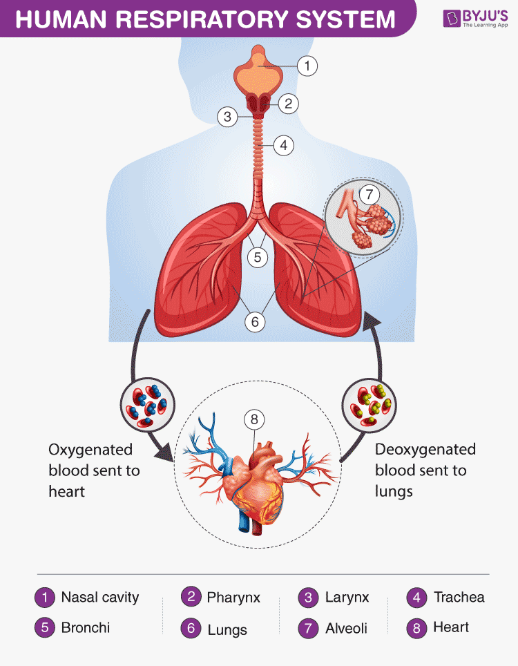
Human Respiratory System Diagram showing different parts of the Respiratory Tract
Features of the Human Respiratory System
The respiratory system in humans has the following important features:
- The energy is generated by the breakdown of glucose molecules in all living cells of the human body.
- Oxygen is inhaled and is transported to various parts and are used in the process of burning food particles (breaking down glucose molecules) at the cellular level in a series of chemical reactions.
- The obtained glucose molecules are used for discharging energy in the form of ATP- (adenosine triphosphate)
Also Read: Difference between trachea and oesophagus
Respiratory System Parts and Functions
Let us have a detailed look at the different parts of the respiratory system and their functions.
Humans have exterior nostrils, which are divided by a framework of cartilaginous structure called the septum. This is the structure that separates the right nostril from the left nostril. Tiny hair follicles that cover the interior lining of nostrils act as the body’s first line of defence against foreign pathogens . Furthermore, they provide additional humidity for inhaled air.
Two cartilaginous chords lay the framework for the larynx. It is found in front of the neck and is responsible for vocals as well as aiding respiration. Hence, it is also informally called the voice box. When food is swallowed, a flap called the epiglottis folds over the top of the windpipe and prevents food from entering into the larynx.
Also check: What is the role of epiglottis and diaphragm in respiration?
The nasal chambers open up into a wide hollow space called the pharynx. It is a common passage for air as well as food. It functions by preventing the entry of food particles into the windpipe. The epiglottis is an elastic cartilage, which serves as a switch between the larynx and the oesophagus by allowing the passage of air into the lungs, and food in the gastrointestinal tract .
Have you ever wondered why we cough when we eat or swallow?
Talking while we eat or swallow may sometimes result in incessant coughing. The reason behind this reaction is the epiglottis. It is forced to open for the air to exit outwards and the food to enter into the windpipe, triggering a cough.
The trachea or the windpipe rises below the larynx and moves down to the neck. The walls of the trachea comprise C-shaped cartilaginous rings which give hardness to the trachea and maintain it by completely expanding. The trachea extends further down into the breastbone and splits into two bronchi, one for each lung.
The trachea splits into two tubes called the bronchi, which enter each lung individually. The bronchi divide into secondary and tertiary bronchioles, and it further branches out into small air-sacs called the alveoli. The alveoli are single-celled sacs of air with thin walls. It facilitates the exchange of oxygen and carbon dioxide molecules into or away from the bloodstream.
Lungs are the primary organs of respiration in humans and other vertebrates. They are located on either side of the heart, in the thoracic cavity of the chest. Anatomically, the lungs are spongy organs with an estimates total surface area between 50 to 75 sq meters. The primary function of the lungs is to facilitate the exchange of gases between the blood and the air. Interestingly, the right lung is quite bigger and heavier than the left lung.
Also Read: Respiration
The respiratory tract in humans is made up of the following parts:
- External nostrils – For the intake of air.
- Nasal chamber – which is lined with hair and mucus to filter the air from dust and dirt.
- Pharynx – It is a passage behind the nasal chamber and serves as the common passageway for both air and food.
- Larynx – Known as the soundbox as it houses the vocal chords, which are paramount in the generation of sound.
- Epiglottis – It is a flap-like structure that covers the glottis and prevents the entry of food into the windpipe.
- Trachea – It is a long tube passing through the mid-thoracic cavity.
- Bronchi – The trachea divides into left and right bronchi.
- Bronchioles – Each bronchus is further divided into finer channels known as bronchioles.
- Alveoli – The bronchioles terminate in balloon-like structures known as the alveoli.
- Lungs – Humans have a pair of lungs, which are sac-like structures and covered by a double-layered membrane known as pleura.
Air is inhaled with the help of nostrils, and in the nasal cavity, the air is cleansed by the fine hair follicles present within them. The cavity also has a group of blood vessels that warm the air. This air then passes to the pharynx, then to the larynx and into the trachea.
The trachea and the bronchi are coated with ciliated epithelial cells and goblet cells (secretory cells) which discharge mucus to moisten the air as it passes through the respiratory tract. It also traps the fine bits of dust or pathogen that escaped the hair in the nasal openings. The motile cilia beat in an ascending motion, such that the mucus and other foreign particles are carried back to the buccal cavity where it may either be coughed out (or swallowed.)
Once the air reaches the bronchus, it moves into the bronchioles, and then into the alveoli.
Respiratory System Functions
The functions of the human respiratory system are as follows:
Inhalation and Exhalation
The respiratory system helps in breathing (also known as pulmonary ventilation.) The air inhaled through the nose moves through the pharynx, larynx, trachea and into the lungs. The air is exhaled back through the same pathway. Changes in the volume and pressure in the lungs aid in pulmonary ventilation.
Exchange of Gases between Lungs and Bloodstream
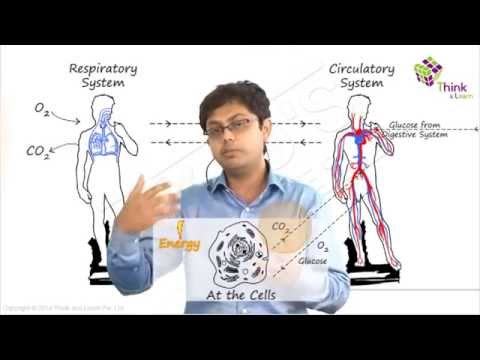
Inside the lungs, the oxygen and carbon dioxide enter and exit respectively through millions of microscopic sacs called alveoli. The inhaled oxygen diffuses into the pulmonary capillaries, binds to haemoglobin and is pumped through the bloodstream. The carbon dioxide from the blood diffuses into the alveoli and is expelled through exhalation.
Also read: Exchange Of Gases in Plants
Exchange of Gases between Bloodstream and Body Tissues
The blood carries the oxygen from the lungs around the body and releases the oxygen when it reaches the capillaries. The oxygen is diffused through the capillary walls into the body tissues. The carbon dioxide also diffuses into the blood and is carried back to the lungs for release.
The Vibration of the Vocal Cords
While speaking, the muscles in the larynx move the arytenoid cartilage. These cartilages push the vocal cords together. During exhalation, when the air passes through the vocal cords, it makes them vibrate and creates sound.
Olfaction or Smelling
During inhalation, when the air enters the nasal cavities, some chemicals present in the air bind to it and activate the receptors of the nervous system on the cilia. The signals are sent to the olfactory bulbs via the brain.
Also Read: Respiratory System Disorders
Respiration is one of the metabolic processes which plays an essential role in all living organisms. However, lower organisms like the unicellular do not “breathe” like humans – intead, they utilise the process of diffusion. Annelids like earthworms have a moist cuticle which helps them in gaseous exchange. Respiration in fish occurs through special organs called gills. Most of the higher organisms possess a pair of lungs for breathing.
Also Read: Amphibolic Pathway
To learn more about respiration, check out the video below:

Frequently Asked Questions
What is the human respiratory system.
The human respiratory system is a system of organs responsible for inhaling oxygen and exhaling carbon dioxide in humans. The important respiratory organs in living beings include- lungs, gills, trachea, and skin.
What are the important respiratory system parts in humans?
The important human respiratory system parts include- Nose, larynx, pharynx, trachea, bronchi and lungs.
What is the respiratory tract made up of?
The respiratory tract is made up of nostrils, nasal chamber, larynx, pharynx, epiglottis, trachea, bronchioles, bronchi, alveoli, and lungs.
What are the main functions of the respiratory system?
The important functions of the respiratory system include- inhalation and exhalation of gases, exchange of gases between bloodstream and lungs, the gaseous exchange between bloodstream and body tissues, olfaction and vibration of vocal cords.
What are the different types of respiration in humans?
The different types of respiration in humans include- internal respiration, external respiration and cellular respiration. Internal respiration includes the exchange of gases between blood and cells, external respiration is the breathing process, whereas cellular respiration is the metabolic reactions taking place in the cells to produce energy.
What are the different stages of aerobic respiration?
Aerobic respiration is the process of breaking down glucose to produce energy. It occurs in the following different stages- glycolysis, pyruvate oxidation, citric acid cycle or Krebs cycle, and electron transport system.
Why do the cells need oxygen?
Our body cells require oxygen to release energy. The oxygen inhaled during respiration is used to break down the food to release energy.
What is the main difference between breathing and respiration in humans?
Breathing is the physical process of inhaling oxygen and exhaling carbon dioxide in and out of our lungs. On the contrary, respiration is the chemical process where oxygen is utilized to break down glucose to generate energy to carry out different cellular processes.
Explore more details about the human respiratory system or other related topics by registering at BYJU’S Biology

Put your understanding of this concept to test by answering a few MCQs. Click ‘Start Quiz’ to begin!
Select the correct answer and click on the “Finish” button Check your score and answers at the end of the quiz
Visit BYJU’S for all Biology related queries and study materials
Your result is as below
Request OTP on Voice Call
| BIOLOGY Related Links | |
Leave a Comment Cancel reply
Your Mobile number and Email id will not be published. Required fields are marked *
Post My Comment
Wow!!!!! Great notes . Thankyou byjus
Wow! this is really helpful. Thank you
Greet notes
Very helpful
Very nice information
good answers!
Thank you so much for the notes
Great notes helpful for students
The notes are really amazing. It helped me a lot. Thank you BYJU’S
The notes are amazing. It helped me a lot. Thank you Byju’s.
Thank you for the notes helpful in the revision
Very good notes. I love Byjus learning application
thank you byjus
These notes are very good for study, thank u so much Byjus.
Helpful for studying. Informative about the topic. Great notes!
Great job you are great Byjus and your notes is superb ❤❤❤❤❣❣❣❣❣🙏🙏🙏🙏
Register with BYJU'S & Download Free PDFs
Register with byju's & watch live videos.

IMAGES
COMMENTS
The respiratory system allows for oxygen to enter the body and carbon dioxide to exit through a series of major organs. Air enters through the nose or mouth and passes through the pharynx, larynx, trachea, bronchi and into the lungs where gas exchange occurs in the alveoli. Oxygen then passes into the bloodstream and carbon dioxide passes out ...
Dipali Harkhani. The respiratory system consists of the nose, pharynx, larynx, trachea, bronchi, bronchioles and lungs. Air enters through the nose and nasal cavity, where it is warmed, filtered and humidified. It then passes through the pharynx, larynx and trachea before entering the lungs via the bronchi. In the lungs, bronchioles divide into ...
The Difference Between Respiration and Breathing. Breathing - Movement of air in and out of the lungs. . Respiration - Also known as cellular respiration, is the energy releasing reaction that takes place inside of cells. Oxygen combines with sugar to release energy.
The Respiratory System
The human respiratory system consists of the upper and lower respiratory tract. The upper respiratory tract includes the nose, nasal cavity, pharynx, larynx, and trachea. The lower respiratory tract includes the bronchi, bronchioles, and lungs. The lungs contain millions of alveoli where gas exchange occurs between inhaled air and blood in the ...
Respiratory System. 2 of 34. Internal Respiration. Internal respiration is the process by which the gases in the air that has already been drawn into the lungs by external respiration are exchanged with gases in the blood/tissues so that carbon dioxide (CO2) is removed from the blood and replaced with oxygen (O2).
Presentation Transcript. The Anatomy and Physiology of the Respiratory System. Functions of the Respiratory System • Air Distribution • Gas exchange • Filters, warms, and humidifies air • Influences speech • Allows for sense of smell. Divisions of the Respiratory System • Upper respiratory tract (outside thorax) • Nose • Nasal ...
35 Anatomy of the Respiratory System Dr. Nbail Khouri MD, MSc. Ph.D. 2 Respiratory System Anatomy. 3 Organization and Functions of the Respiratory System. Consists of an upper respiratory tract (nose to larynx) and a lower respiratory tract ( trachea onwards) . Conducting portion transports air. - includes the nose, nasal cavity, pharynx ...
2. Larynx - part of the trachea where our vocal cords are located. 3. Trachea - the tube that leads from the nose and mouth to the lungs. The walls have rings of cartilage to protect the trachea and prevent it from collapsing. The Respiratory System How we get our oxygen and get rid of carbon dioxide. HTML view of the presentation.
This 69 slide power point presentation will help your students to learn about the Respiratory system. Topics include: anatomy/function of respiratory system, the respiration process, gas transport, respiratory volumes and capacities, and factors affecting respiratory rate. Ideal for an Anatomy and Physiology or related course and can be used in ...
Activity of respiratory muscles is transmitted to the brain by the phrenic and intercostal nerves Neural centers that control rate and depth are located in the medulla The pons appears to smooth out respiratory rate Normal respiratory rate (eupnea) is 12-15 respirations per minute Hypernia is increased respiratory rate often due to extra ...
The respiratory system has several key functions including supplying oxygen to the body and removing carbon dioxide. It consists of breathing, which has two phases - inhalation that draws air into the lungs and exhalation that forces air out. Gas exchange occurs externally between the lungs and blood and internally between blood and tissues.
Four Respiration Processes. Breathing (ventilation): air in to and out of lungs. External respiration: gas exchange between air and blood. Internal respiration: gas exchange between blood and tissues. Cellular respiration: oxygen use to produce ATP, carbon dioxide as waste.
Respiratory system.ppt - Free download as Powerpoint Presentation (.ppt), PDF File (.pdf), Text File (.txt) or view presentation slides online. The respiratory system has several functions: (1) It allows for the exchange of gases between the atmosphere and body cells through the processes of respiration and breathing. (2) It involves both upper and lower respiratory tract organs including the ...
Presentation on theme: "Respiratory Physiology"— Presentation transcript: 3 1. Pulmonary Ventilation. 4 2. External Respiration Gas exchange (oxygen loading and CO2 unloading) between the pulmonary blood and alveoli EXternal respiration = gas exchange between the blood and body EXterior. 5 3. Respiratory Gas Transport. 6 4.
What is a Respiratory Surface? • In all organisms, the exchange of gases must occur across a respiratory surface. • Must be moist • Must be very thin so that gases are able to pass through • Must be a supply of oxygen • Must be closely connected to the transport system to deliver gases to and from cells. The Human Respiratory System.
The respiratory system collects oxygen and releases carbon dioxide. Air enters through the nose and mouth then travels through the trachea and bronchi, which divide into bronchioles ending in alveoli. The alveoli are surrounded by capillaries that exchange oxygen and carbon dioxide through the thin alveolar walls. Oxygen is absorbed into the ...
610 likes | 666 Views. Assessment of respiratory system. Dr .Isazadehfar Specialist in community and preventive medicine. Anatomy and physiology. The respiratory tract extends from the nose to the alveoli and includes not only the air-conducting passages but also the blood supply. Download Presentation.
It travels down the oesophagus and down the bronchi and bronchioles. 4. The lungs fill up with air increasing the volume and making the rib cage move upwards and outwards. 5. The diaphragm contracts and fattens. 6. The air moves from the bronchioles and into the alveoli where oxygen diffuses into the blood. 7.
Explore the Respiratory System learning module on AccessMedicine. AccessMedicine is a subscription-based resource from McGraw Hill that features trusted medical content from the best minds in medicine.
The respiratory system has several key functions including supplying oxygen to the body and removing carbon dioxide. It consists of breathing, which has two phases - inhalation that draws air into the lungs and exhalation that forces air out. Gas exchange occurs externally between the lungs and blood and internally between blood and tissues.
The respiratory tract in humans is made up of the following parts: External nostrils - For the intake of air.; Nasal chamber - which is lined with hair and mucus to filter the air from dust and dirt.; Pharynx - It is a passage behind the nasal chamber and serves as the common passageway for both air and food.; Larynx - Known as the soundbox as it houses the vocal chords, which are ...
The document summarizes the examination of the respiratory system. It describes inspecting the chest shape and movements, palpating the apex beat and trachea position, percussing the chest to compare resonance, and auscultating breath sounds including vesicular, bronchial, vocal resonance, and added sounds like rhonchi or crepitations. The ...